

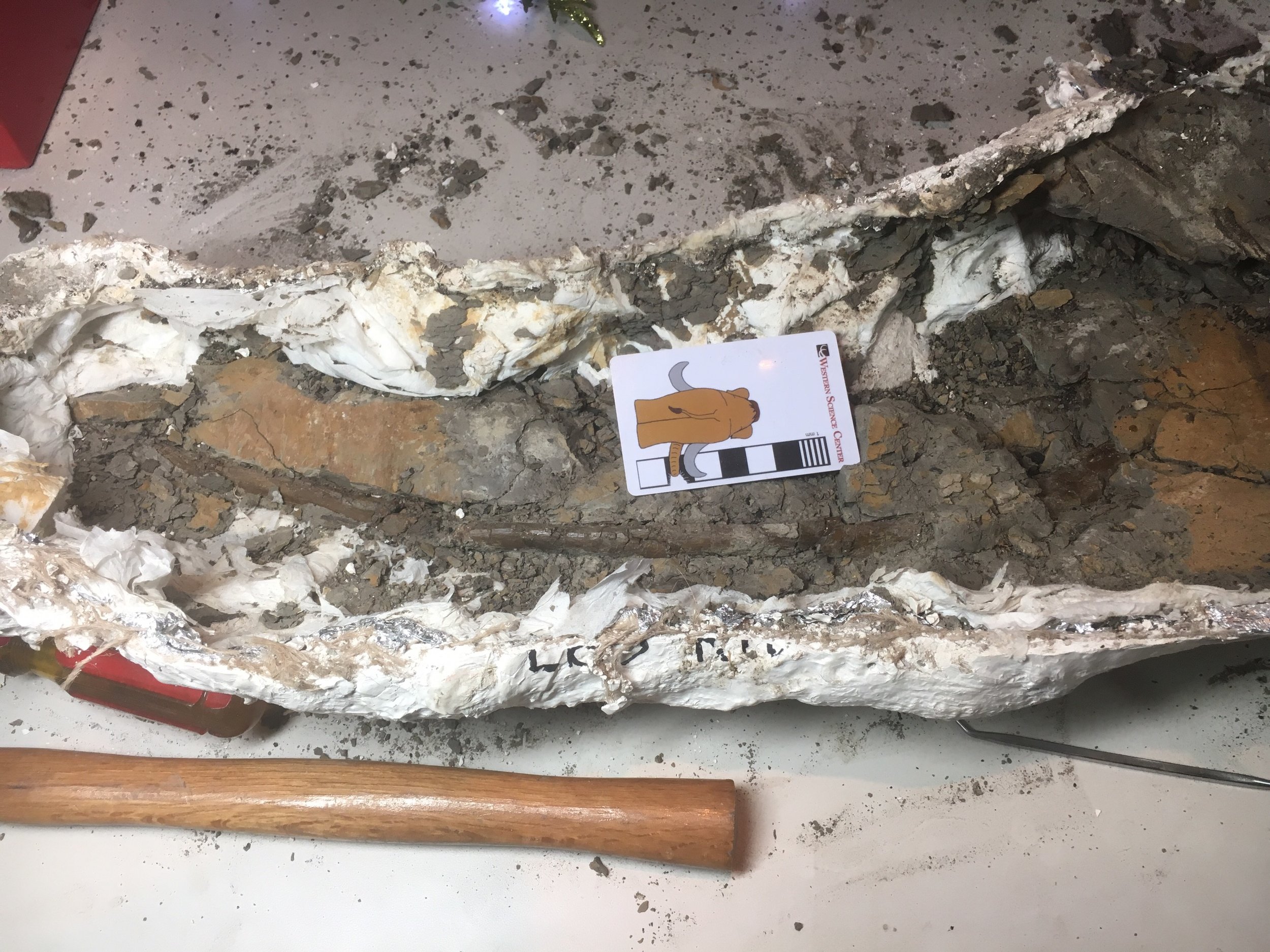
 A few months ago Dr. Andrew McDonal joined the staff of WSC as our new curator. Andrew has been excavating for several years in the Cretaceous Menefee Formation on federal land in New Mexico, and now some of that material is making its way to WSC. This morning we opened our first Menefee Formation field jacket on public view at the museum’s Exploration Station. The image above shows the jacket after about 3 hours of prep work. The jacket contains a ceratopsian rib, the posterior edge of which is visible running through the middle of the jacket from the left end to about 10 cm past the top of the scale bar. The rib actually continues all the way to the other end of the jacket, but we’ve not yet uncovered it that far. The small black specks dotting the sediment are fragments of charcoal from burned Cretaceous trees.Andrew tells me that this rib is part of an associated skeleton including at least a dozen bones, from a small, probably juvenile, ceratopsian. So far we don’t know what taxon it represents, but there’s still a lot of material to prepare. This rib will be on public view in the Exploration Station for the next few weeks as we continue our preparation work.
Fossil Friday - printed bison calcaneum
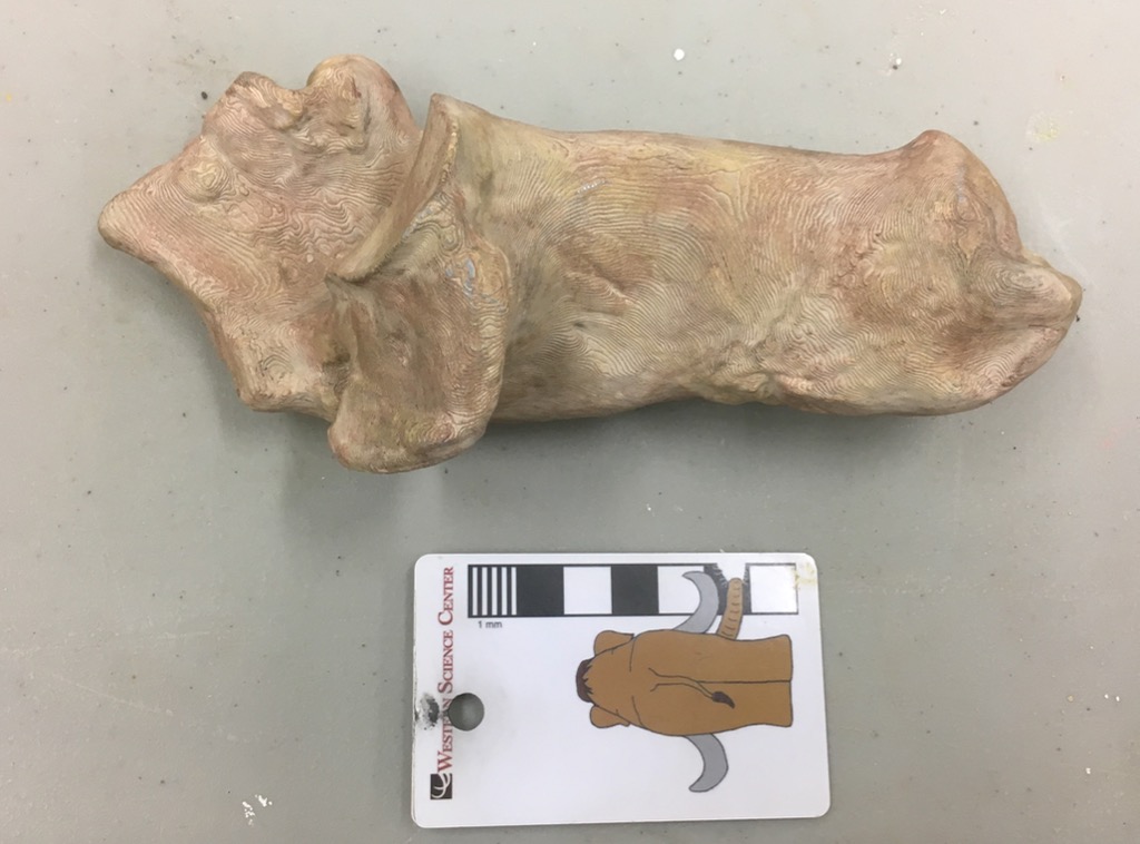 At our annual Science Under the Stars fundraiser last September, or donors provided us with funding to start a #D scanning and printing lab the the Western Science Center. While some of our equipment is still on order, our first printer arrived a few weeks ago and we've been printing as much as possible while we train ourselves in its use. During the Valley of the Mastodons Symposium, Bernard Means from the Virtual Curation Lab scanned a number of our specimens. While we wait for our scanner to arrive we've been printing some of Bernard's scans.Earlier this week we printed a right Bison calcaneum, seen above in medial view. The original specimen is exhibited in the floor case with the mastodon "Little Stevie" (they were found together). It's difficult to photograph through this case (below), and it's difficult to open; the symposium is the first time it has been opened in 11 years! So while the case was opened we scanned as much as possible, including this calcaneum (which is visible in the center of the case photo below):
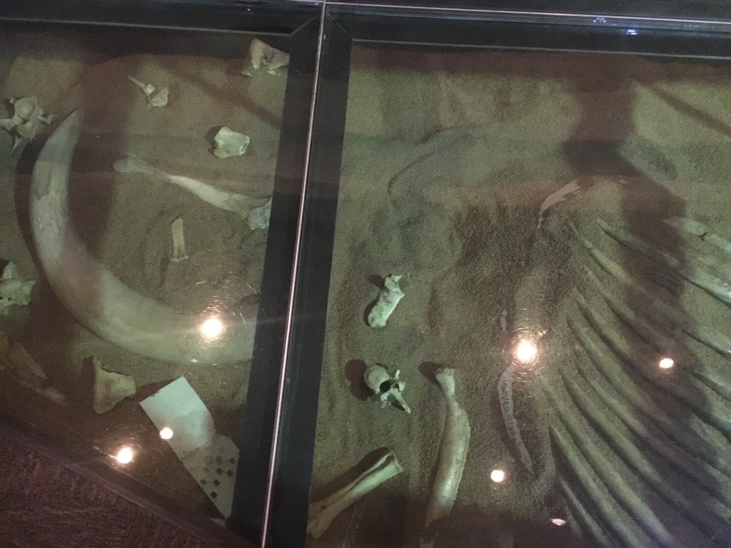 This bone was our largest single print to date, and took about 24 hours to print. We've since printed a slightly larger bone as we continue to explore the limits of the printer.
 The calcaneum is part of the foot and ankle structure, and forms the heel and the attachment point for the Achilles tendon. This particular specimen was collected at Diamond Valley Lake's East Dam, and is roughly 45,000 years old. The original specimen is now back on display in its floor case, while the printed replica will be added to our teaching collection.
Fossil Friday - deer humerus
 One of the collections at WSC comes from the Harveston neighborhood of Murrieta in Riverside County. While this is a fairly small collection, it’s amazingly diverse, with well over a dozen different species of mammals. A group of us have started going through this collection to document it. One of the Harveston specimens is a small deer, probably the genus Odocoileus. The element above is the distal end of the humerus. Modern Odocoileus includes the mule deer O. hemionius and the smaller white-tailed deer O. virginianus. Interestingly, the Harveston specimen is quite a bit smaller than modern white-tailed deer, even though it’s an adult. We have a few other deer elements from the Harveston material that we’ll have to compare to see if the size difference is consistent.
Fossil Friday - printed mastodon molar
 Each September, Western Science Center holds an annual fundraiser called Science Under the Stars to raise funds for museum operations. At the end of the evening we often do a final “Special Ask” to raise dedicated funds for a particular project. This year, for the Special Ask we requested funds for a 3D-scanning, photogrammetry, and printing lab. Our donors, led by the Soboba Band of Luiseño Indians, came through in spectacular fashion.
  Our first purchase for the lab, a LulzBot TAZ6 printer (above), arrived last week. After our initial demo print to make sure everything was working, our first specimen print was a mastodon tooth (of course). It was printed from a digitial file provided to us by VCU’s Virtual Curation Lab, courtesy of Bernard Means and Terrie Simmons-Ehrhardt, which was in turn based on CT scans provided by California Imaging and Diagnostics. The particular tooth is the lower right 3rd molar of a West Dam mastodon we refer to as The Outlier, so named because its 3rd molars are the widest ones known from California (although still narrower than the average m3 from the rest of the country).In a few weeks we’ll be purchasing our scanning and photogrammetry equipment, as well as additional printers, so you’ll be hearing a lot more about this in the months ahead. Next week I’m attending the Geological Society of America meeting, where Brett and I will be presenting on both the “Stepping Out of the Past” and the “Valley of the Mastodons” exhibits, and I’ll have updates from there as well.
Fossil Friday - vole tooth
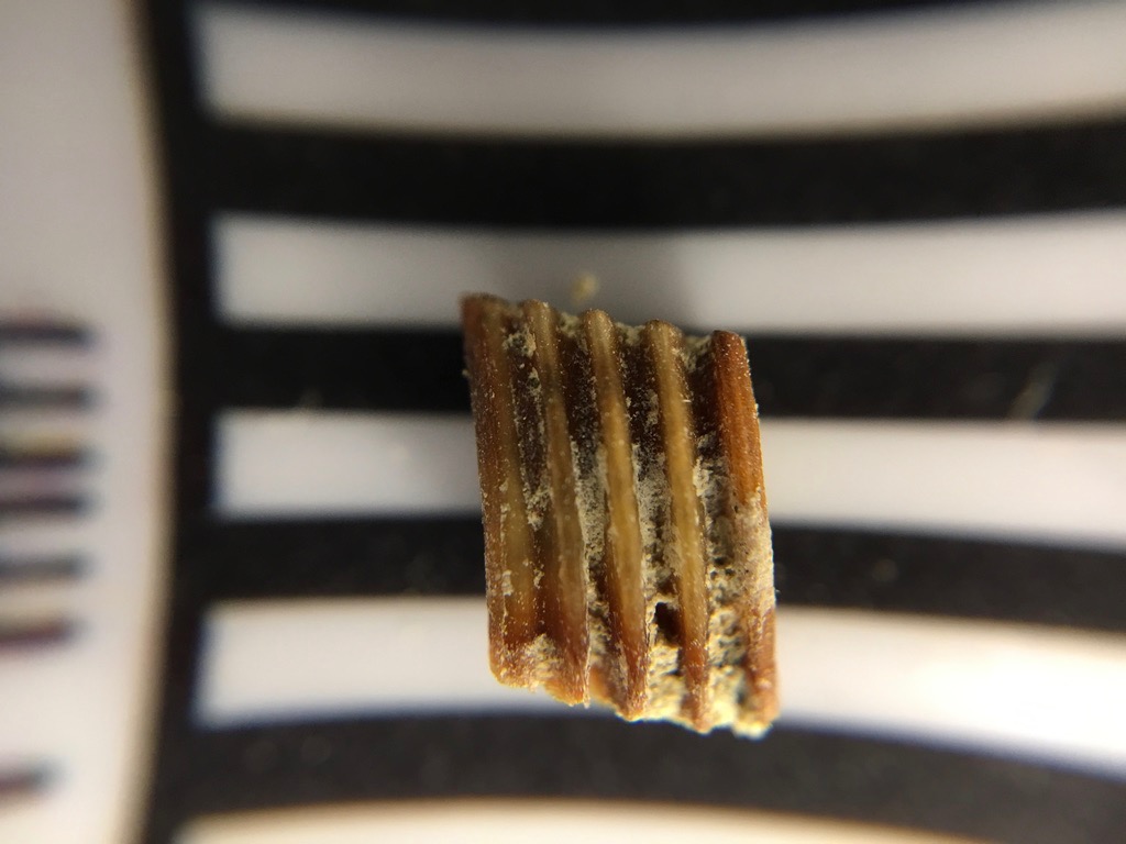 This week is National Rodent Awareness Week. Fossil rodents may not get a lot of headlines, but they are often the most common vertebrate fossils in Neogene terrestrial deposits, and have the potential to convey huge amounts of information about age and paleoenvironment.One of the larger collections at the Western Science Center is from the Southern California Edison El Casco substation in Riverside County. These Late Pliocene/Early Pleistocene deposits of the San Timoteo Formation produced over 16,000 fossils, including large numbers of rodents.Shown above is the right lower first molar of Microtus, a vole. It's shown in medial view, with anterior to the left. The black and white bands under the tooth are each 1 mm thick, so the whole tooth is about 4 mm tall.Voles and other members of the subfamily Arvicolinae (including hamsters, lemmings, and muskrats) are herbivores, and often eat abrasive food such as grasses. Like many other herbivores, this is reflected in their tooth anatomy, which includes loops of enamel with exposed dentine in the middle. Because the enamel is harder than the dentine, it always sticks up a bit higher than the dentine and allows the tooth to stay sharp even as it wears down. The enamel loops are expressed as ridges or columns when seen from the side, and are clearly visible in the view above. In occlusal view (below), the loops are expressed as a series of alternating triangles, a pattern that is characteristic of the arvicolines.
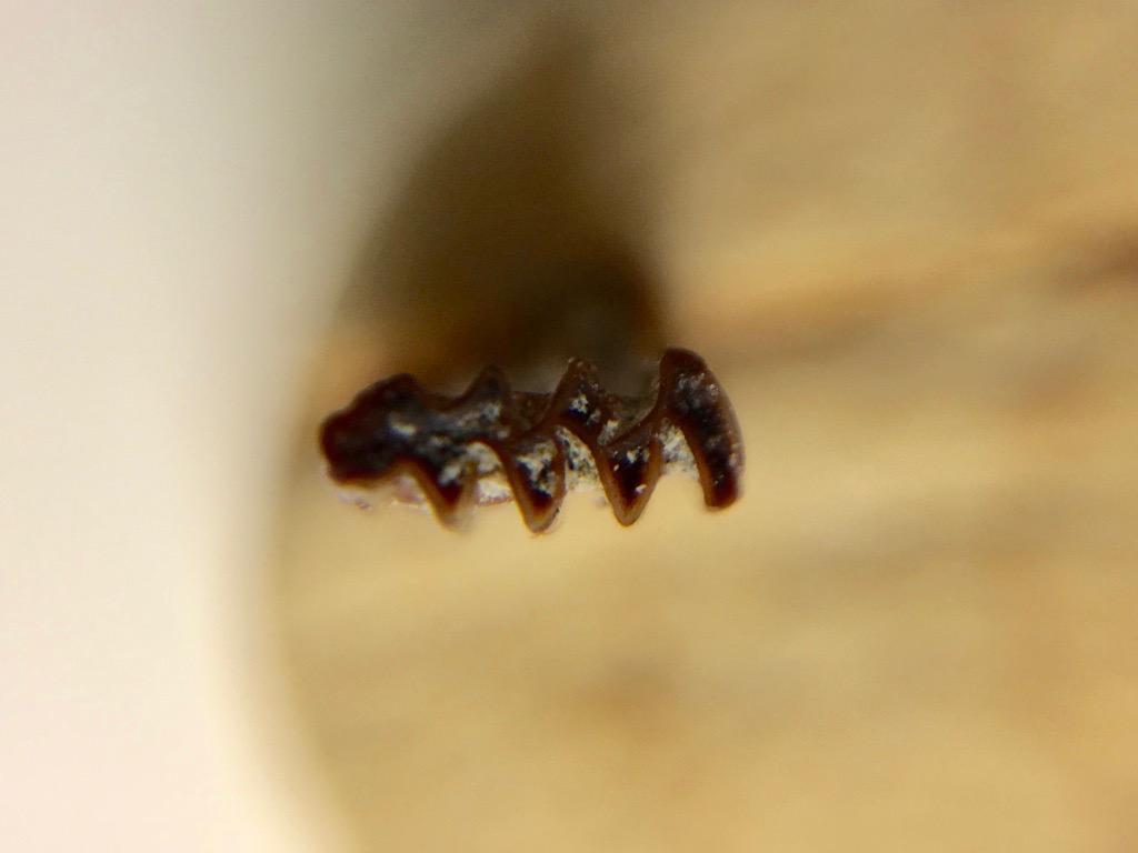
Fossil Friday - cat skull fragments
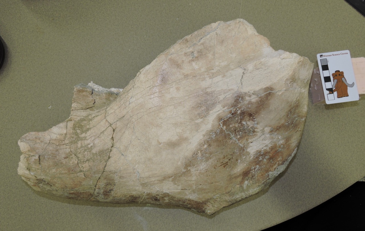
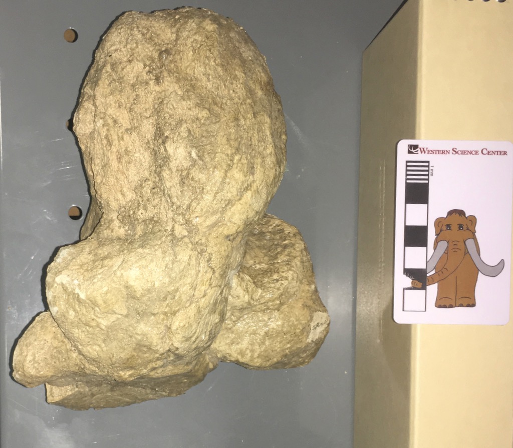
 With many fossils, particularly vertebrates, small fragments can sometimes be very informative and justify close scrutiny. That was the case with a pair of associated bones from Diamond Valley Lake. One of the fragments, shown above, includes the occipital condyles that form the articulation between the skull and the first vertebra. The condyles sit on either side of the foramen magnum, the opening through which the spinal cord passes.The second fragment, below, appears to be part of the bottom of the skull just anterior to the condyles, including portions of the left squamosal and basisphenoid.
 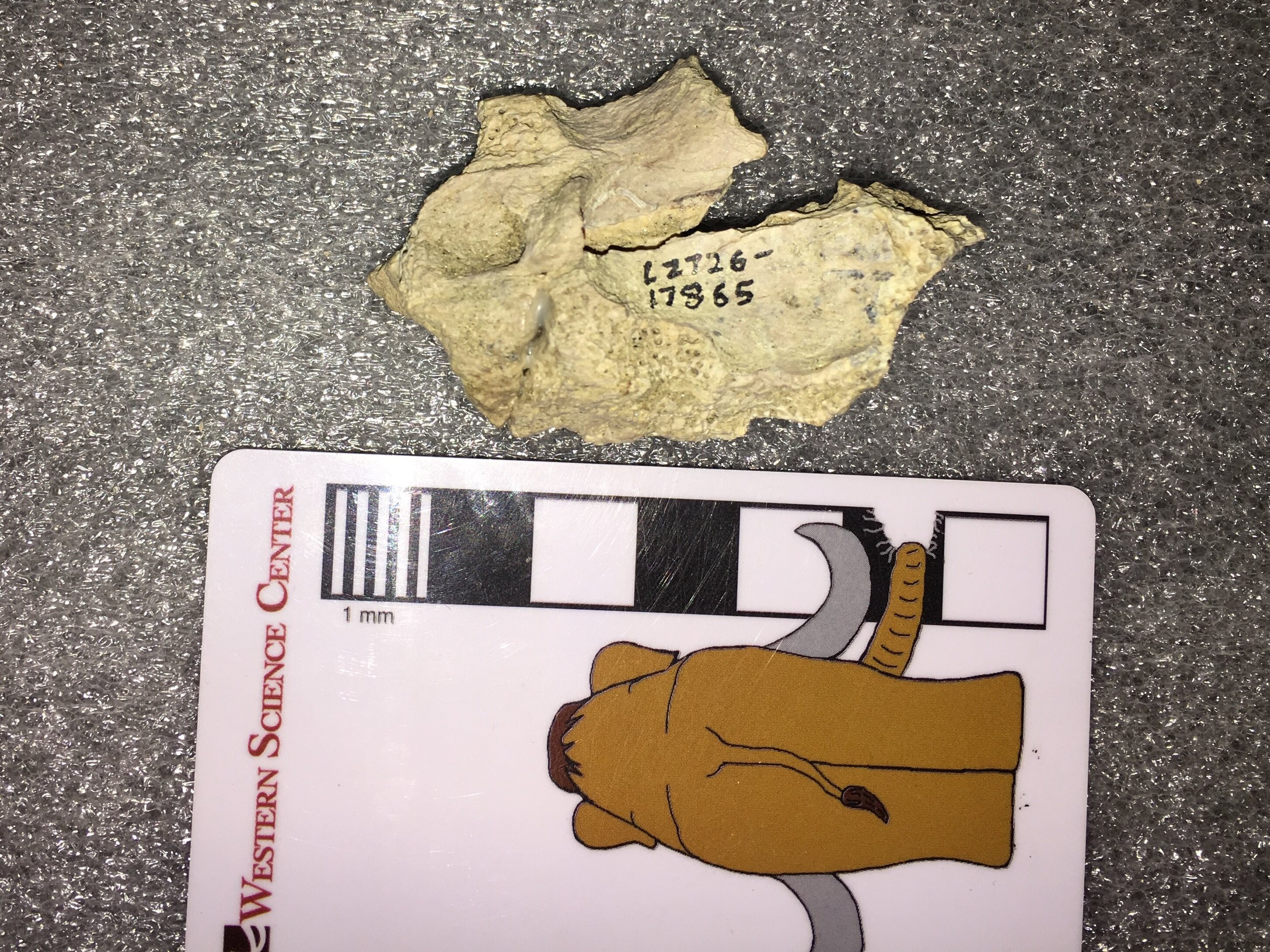 The initial DVL report identified these fragments as coming from a cat, but the type of cat was uncertain. They appear to be far too large to come from a mountain lion, and the report suggests they may be from a jaguar, Panthera onca. Today jaguars are only found in South America and as far north as Mexico (except for one individual roaming the mountains near Tucson, Arizona), but the species originated in North America and was widespread in the southern US states during the Pleistocene. However, as pointed out in the DVL report, it was very rare in the Pleistocene of Southern California and would be a surprising find at Diamond Valley Lake. We have a cast jaguar skull in our teaching collection that allows for comparison:
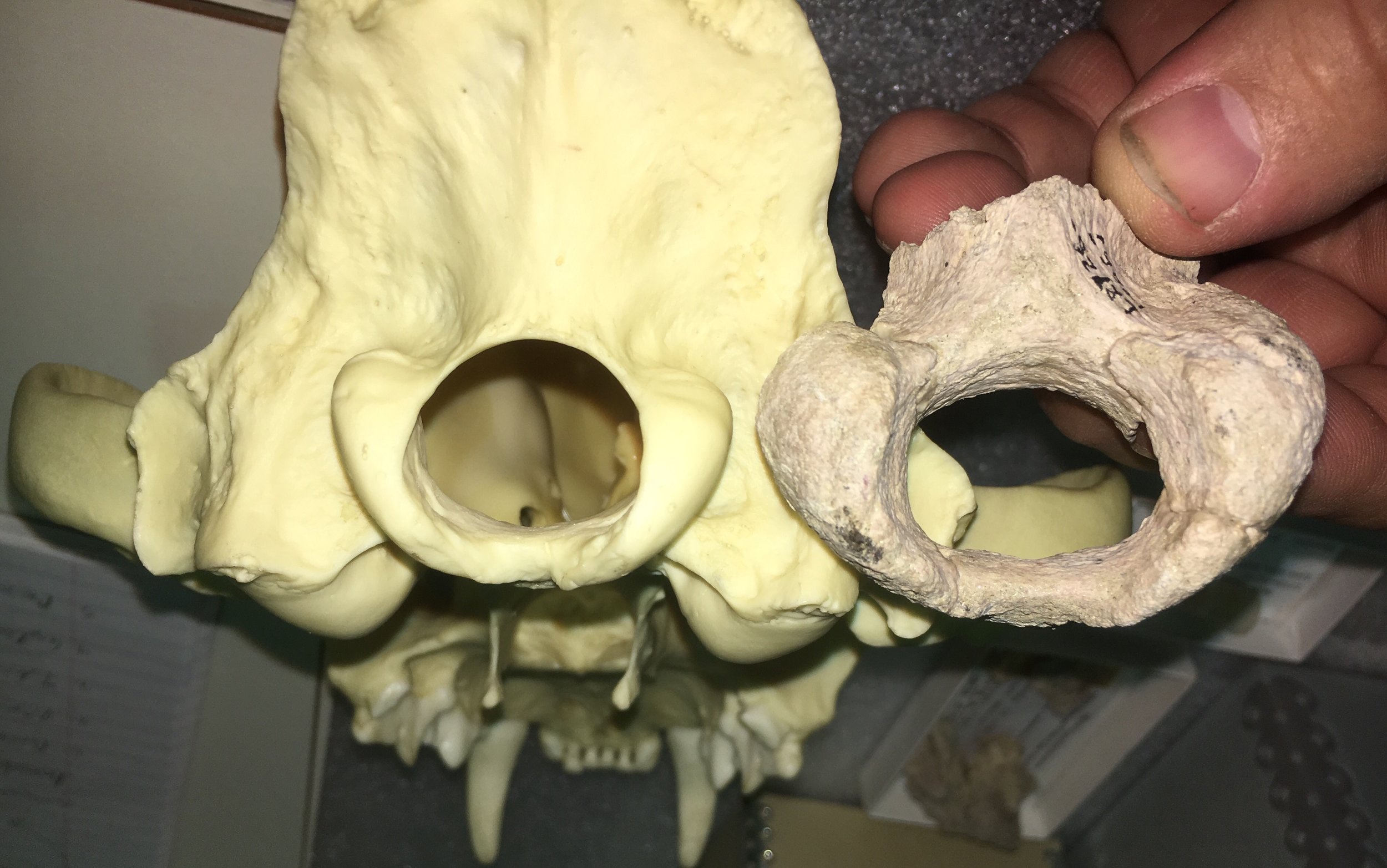 The DVL specimen is much larger than the modern cast, but that's not a problem; Pleistocene jaguars were larger than modern ones. The shape is somewhat similar, but there are differences in the shape of the condyles. In an oblique ventral view (below) it's also clear that the basioccipital (the bone just in front of the condyles) is shaped quite differently:
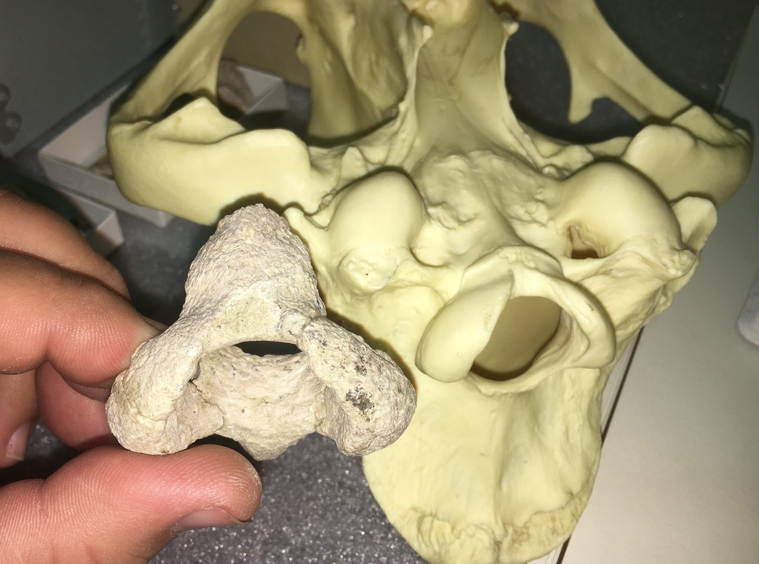 As it turns out, this fragment is a pretty close (if not perfect) match in size and shape for our cast of Smilodon from Rancholabrean La Brea:
 Turning to the squamosal, below is the relevant area on our cast Smilodon; note that an additional bone, the tympanic bulla, is cast into this specimen and obscures part of the squamosal:
 Here's the same area with the DVL squamosal superimposed:
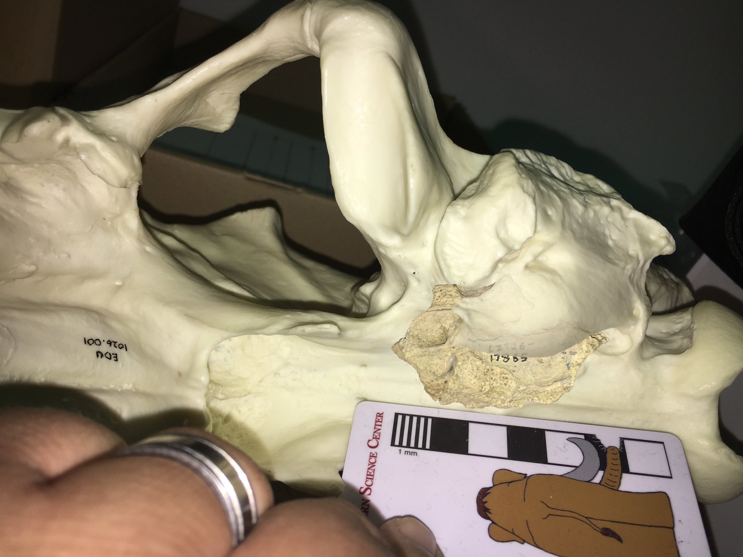 This appears to be a very close match. There's no single feature that definitively places these bones in Smilodon, but it seems to be a closer match than Panthera onca. Moreover, while jaguars are rare in Southern California and otherwise unknown from Diamond Valley Lake, sabertooth cats are very common in Southern California deposits and are known from several DVL sites. Taken together, I think everything points to this specimen being Smilodon.* Edited to include the correct the range of the modern jaguar.
Fossil Friday - Bison molar
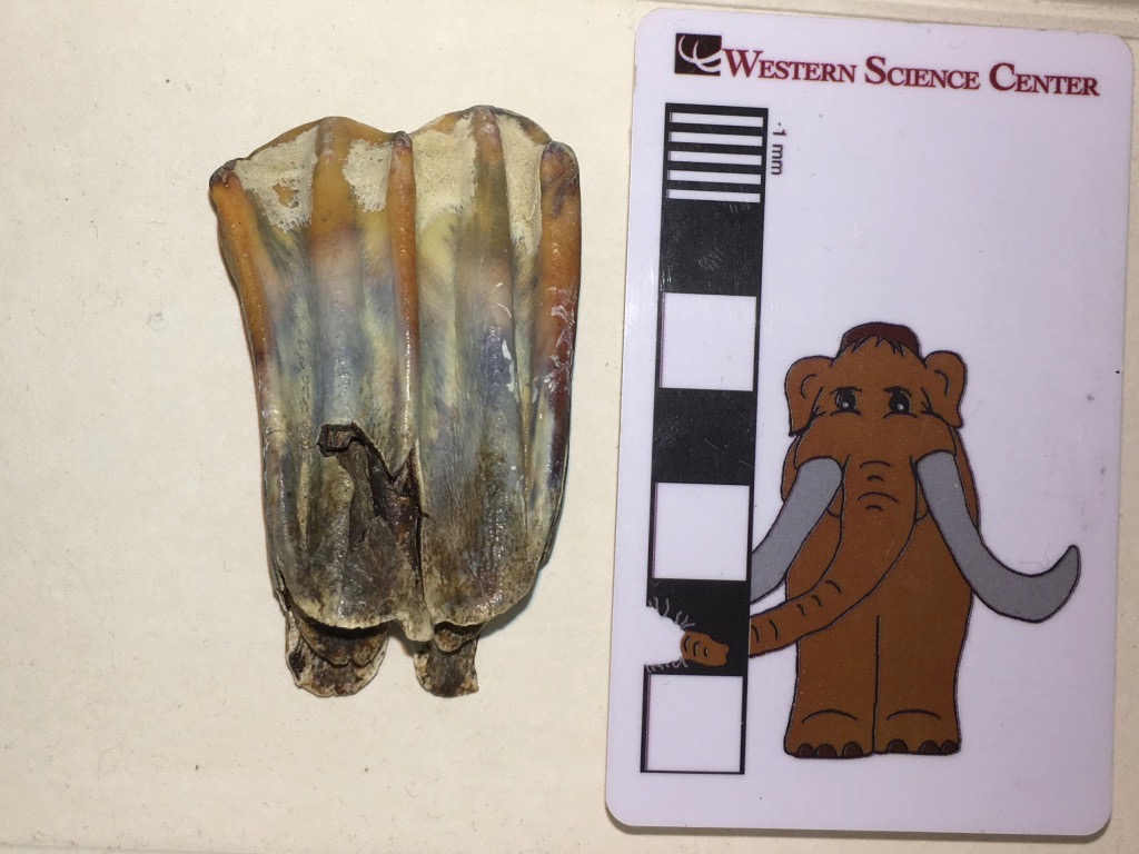 At Diamond Valley Lake, five genera account for 93% of the preserved large animals: Bison, Equus, Camelops, Mammoth, and Paramylodon. Of these five, the most abundant are Bison.The tooth shown above is an upper left 1st molar from a bison. Unfortunately, I photographed it upside down, so even though it's an upper tooth in the image the edge at the top is the chewing surface and the roots are at the bottom. This is the labial view, the side of the tooth closest to the lips.Below is the lingual view, showing the side of the tooth closest to the tongue:
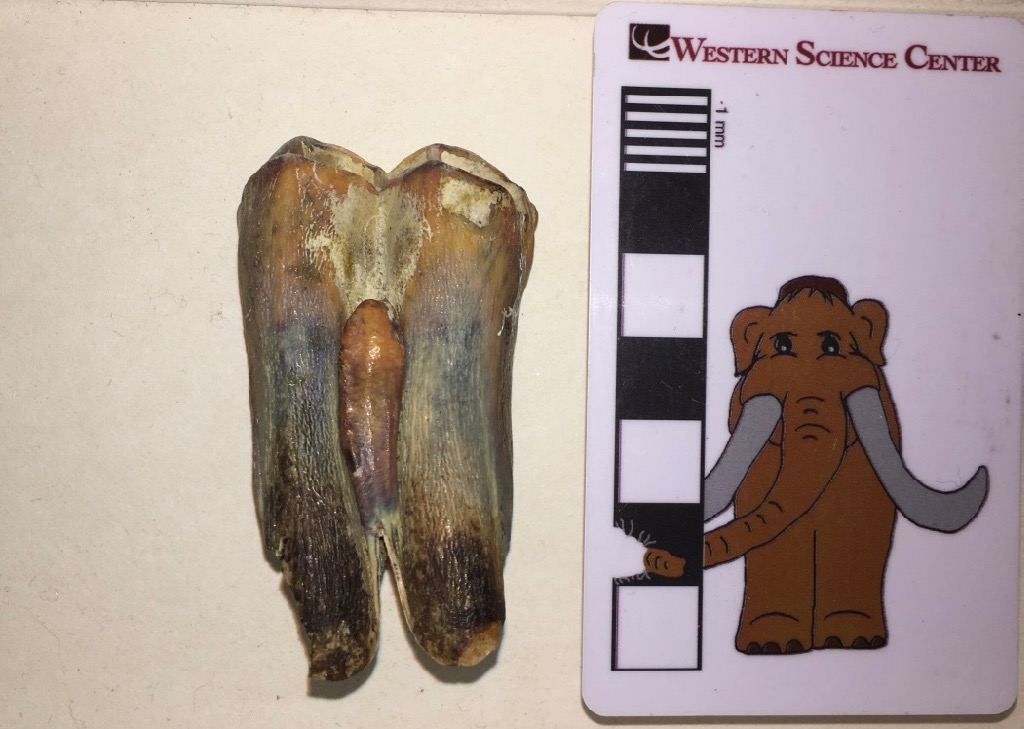 Again, the occlusal edge is at the top, roots at the bottom. The reddish-brown protuberance in the middle of the tooth is a column called the style (stylid on the lower teeth). The style is almost always present in the upper and lower molars of Bison, but it's very rare in the closely related cattle genus Bos. So it's a very convenient identification feature (if not necessarily 100% reliable) for distinguishing between Bos and Bison.In occlusal view (below), the style can be seen end-on on the lingual side of the tooth (on the left in this image):
 This particular tooth only has very light wear. In a heavily-worn molar, the tooth eventually wears away so much that the occlusal surface of the tooth is level with the tip of the style. The style then begins to wear as well, and becomes visible on the occlusal surface as an isolated ring of enamel. Because the style is not the same shape throughout its length, as wear continues the ring gradually becomes an oval, and then a loop of enamel connected to the rest of the tooth. So the style shape on the occlusal surface give a rapid indication of how worn the tooth is, even is it's still embedded in the socket.This tooth was collected from the West Dam of Diamond Valley Lake. Bison latifrons has not been identified from the West Dam, so this is most likely Bison antiquus. Western Science Center also has teaching kits based on interpretation of bison tooth wear in cast specimens available for purchase.
Fossil Friday - mastodon vertebra revisited
 After a highly successful Science Under the Stars fundraiser, I've tried to get back into the lab to catch up on neglected science work. Administrative duties are still conspiring to keep me chained to the phone and computer, but I have made some progress.Above is a mastodon thoracic vertebrae, one of the same vertebrae featured on Fossil Friday last month. We've removed this vertebra from the field jacket and started repairs and reconstruction.The image above is in posterior view, with anterior to the top. This is an anterior thoracic, with the very tall neural spine typical of that part of the skeleton. There are two prominent articulations on the right side for ribs. One is just below the tip of the transverse process and faces toward the lower right, while the other is the circular depression between the transverse process and the centrum. These two regions actually articulate with different ribs; the transverse process articulates with the tuberculum (or tubercle) of the rib, while the depression adjacent to the centrum articulates with the capitulum (or head) of the next rib.We'll continue cleaning this and the other bones from this jacket in the coming weeks. We should be able to pin down pretty tightly which vertebra in the series this is. Then these will be joining other bones from the same individual in the Valley of the Mastodons exhibit.
Fossil Friday - mammoth tooth
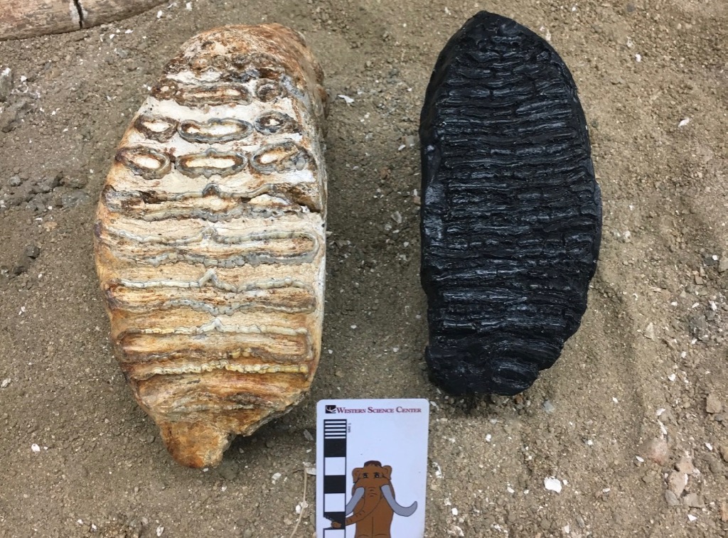 Tomorrow is the Western Science Center's annual fundraiser, Science Under the Stars. That has our entire staff, including me, wrapped up in preparations for 500 guests and a massive benefit auction, so Fossil Friday will necessarily be brief.Above on the left is a partial molar from a Columbian mammoth, Mammuthus columbi, from Pleistocene deposits near Murrieta in Riverside County. As massive as the tooth is, it's incomplete, with the anterior part of the tooth having already worn away. Mammoth teeth (in fact, all the elephants) have enamel arranged in transverse plates that wear to enamel loops on the occlusal surface.The tooth on the right is a cast of a wooly mammoth (M. primigenius) from Alaska. While it's a little difficult to see on the black tooth, the enamel plates on the wooly mammoth are thinner and more numerous than on the Columbian mammoth. This general rule is often used to distinguish between the two species, but as so often seems to be the case, proboscideans are not good at following rules! There is considerable overlap in this character ("lamellar frequency") between Columbian and wooly mammoths. Moreover, it appears that the two species were regularly interbreeding where their ranges overlapped (the apparent hybrids were originally given a different species name, M. jeffersonii). And recent genetic evidence calls into question whether Columbian and wooly mammoths were in fact just different morphs of a single, highly variable species.
Fossil Friday - mammoth tusk
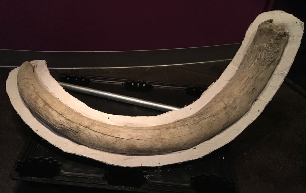 I missed last Fossil Friday while I was attending the Society of Vertebrate Paleontology in Calgary, which included posters on Diamond Valley Lake mastodon tusks and the Valley of the Mastodons Symposium and Exhibit. But I'm now back in Hemet, and ready for some more Fossil Friday posts!A few months ago I posted about a Columbian mammoth lower jaw from Merced County, collected by PaleoResources during a Caltrans road project. There was actually quite a bit of material recovered during this project, and we're currently in the process of integrating it into the WSC collections. One of the other large pieces is the tusk shown above.This is a typical left tusk from a mammoth, with strong curvature in multiple planes resulting in an open spiral shape. Mastodon tusks, in contrast, tend to be thicker for a given length (at least in males), and curve more strongly in one plane than the other, so the tusk is more of an arc than a spiral. While it's difficult to see from this angle, the tusk shown here has a fairly prominent wear facet at the tip.When PaleoResources dropped off this collection, they indicated that they suspected the mammoth material represented more than one individual, even though there were no duplicate pieces (so that, technically, the minimum number of individuals = 1). I agree with PaleoResources' assessment; I don't see any way such a large tusk could have come from a mammoth as young as the one represented by the lower jaw in the same deposit.This tusk, along with the juvenile lower jaw and an associated mammoth pelvis, are currently on display at the Western Science Center.
Fossil Friday - mastodon vertebrae
 Even though the Valley of the Mastodons exhibit is now open, it doesn't mean that our mastodon work has moved to the backburner. On the contrary, we're now doing more mastodon work than ever before!The field jacket shown above is associated with a partial mastodon skeleton currently on display in Valley of the Mastodons; I've previously featured the axis vertebra and the tusks from this mastodon on Fossil Fridays. This jacket has been partially prepared, but there are still several bones present. Some of these are highlighted in the color-coded version below:
 The blue area is (I think) the first cervical vertebra, or atlas. Yellow is a rib, and red are two thoracic vertebrae. There are also several unmarked and unidentified fragments. The thoracic vertebrae have the very tall neural spines that are typical of anterior thoracics in animals with large, heavy heads.We'll be removing these bones over the next few weeks, and as they're completed they'll be moving into the Valley of the Mastodons exhibit along with the rest of this mastodon.Next week I'll be in Calgary, attending the Society of Vertebrate Paleontology annual meeting. Many paleontologists will be live-tweeting the conference, including me (at least on occasion) and @MaxMastodon.
Fossil Friday - Aviculopecten
 During last week's Valley of the Mastodons events, museum supporter Doug Shore donated a collection of invertebrate and plant fossils to Western Science Center. Several of Doug's specimens come from Mason Creek, an incredibly rich Carboniferous site in Illinois that is most famous for its beautiful fossil plants. While WSC has a large collection of Mason Creek plants, we did not have any of the numerous animals also found at the site until now.Above are two specimens of the mollusk Aviculopecten mazonensis, a scallop. At least, I think it's two specimens. As the name states, bivalve mollusk have two valves or shells. In many bivalves these two shells look quite different from each other, but in the pectinids (scallops) they are often very similar (although one valve tends to be deeper than the other). In this case the two valves have slightly different dimensions and both seem to have the same curvature, making me suspect they represent two individuals rather than two shells from one individual. Thanks to Doug for this nice addition to the WSC collection!
Fossil Friday - mastodon tusks
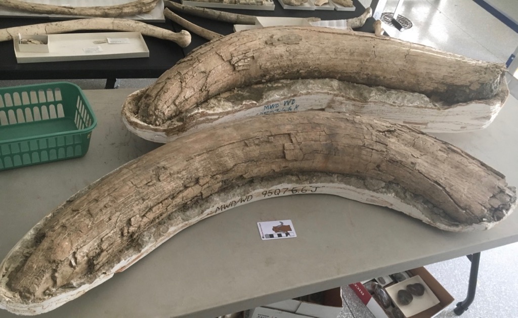 In three days scientists start arriving in Hemet for the Valley of the Mastodons symposium, and we're one week from the opening of the associated Valley of the Mastodons exhibit. As a result, I'm a little swamped, and today's Fossil Friday post will have to be brief.The original plan for Valley of the Mastodons was to display all of the mastodon material recovered from Diamond Valley Lake. That plan didn't last long. Our temporary exhibit gallery is 3,000 square feet, and while that's a pretty big room, we have a LOT of mastodons! It didn't take long to realize that they won't all fit. So we had to triage the collection, and things that have really cruddy preservation, and isolated bones that we can't say much about, are not going on display.One of the specimens that did make the cut is a partial mastodon from the West Dam, the area with the highest concentration of mastodons. This partial skeleton includes several ribs and vertebrae (a few weeks ago I featured its axis vertebra), a lower jaw fragment, a humerus, and both tusks. The tusks are shown above. They're reasonably well preserved, and include the open pulp cavities at the proximal ends (filled with sediment). The tips had crumbled a bit and are incomplete, but it doesn't look like too much is missing. The size of these tusks indicate that this was an adult male mastodon, although the unfused epiphyses on several vertebrae show that it wasn't quite finished growing.This mastodon and more than a dozen others will be on display beginning August 5 (WSC members are invited to a preview showing on August 4).
Fossil Friday - mastodon jaw fragment
 As we build up to our new exhibit opening, I'll continue the Month of Mastodons with a partial lower jaw.This is the back end of the left dentary, seen in lateral view with anterior to the left. The front half of the bone is missing, as are the coronoid process and condyle that should be located at the posterodorsal edge (upper right in the photo). The tooth crown is also broken. Below is a medial view of the same bone:
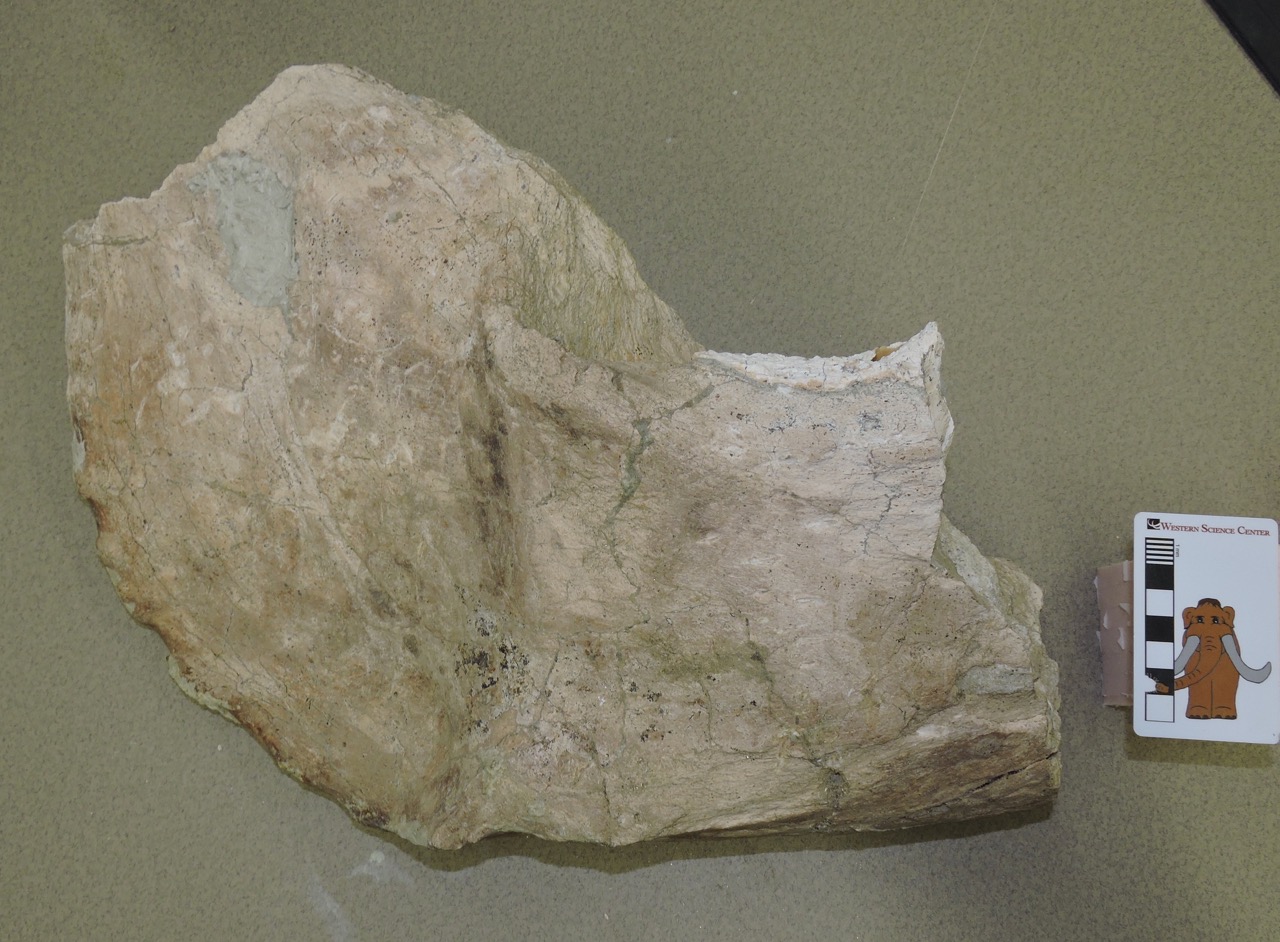 And here's the dorsal view:
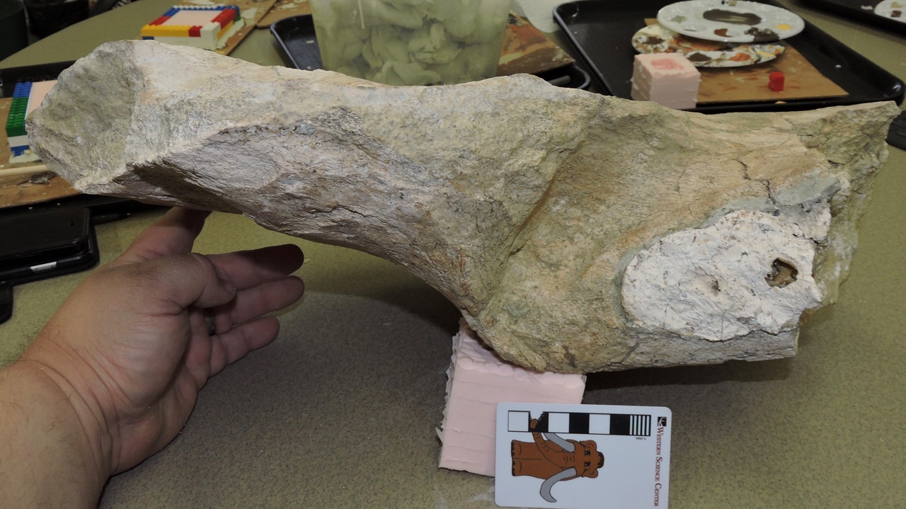 In this view the broken tooth is more clearly visible. Even though the crown is broken off, the distinctive mastodon tooth shape is clear.Unlike the vast majority of mastodons in the Western Science Center collections, this jaw is not from Diamond Valley Lake. Instead, it was collected from the Grizzly Ridge section of Murrieta, about 20 km southwest of Diamond Valley Lake (still in Riverside County). This specimen came in as part of a mitigation project, and unfortunately included almost no data except for the locality name. Both early and late Pleistocene deposits are known from Murrieta, so we're not yet sure of the age.We have not yet begun preparation of this specimen, but a quick survey indicates that a substantial part of the skeleton was recovered, including at least some vertebrae, numerous ribs, multiple limb elements, and several tusk fragments. We're hoping to prepare the specimen over the next year or so in our new Exploration Station prep area in our main exhibit gallery. The dentary fragment will also be on exhibit during Valley of the Mastodons, opening to the public on August 5.
Fossil Friday - mastodon partial skull
 Our latest mastodon installment as we approach the "Valley of the Mastodons" exhibit opening 3 weeks from now is "Braces", a partial mastodon skull from the San Diego Canal."Braces" is still in his field jacket, with the ventral side exposed (and the large tusks do indicate that he's male). Below is an annotated version:
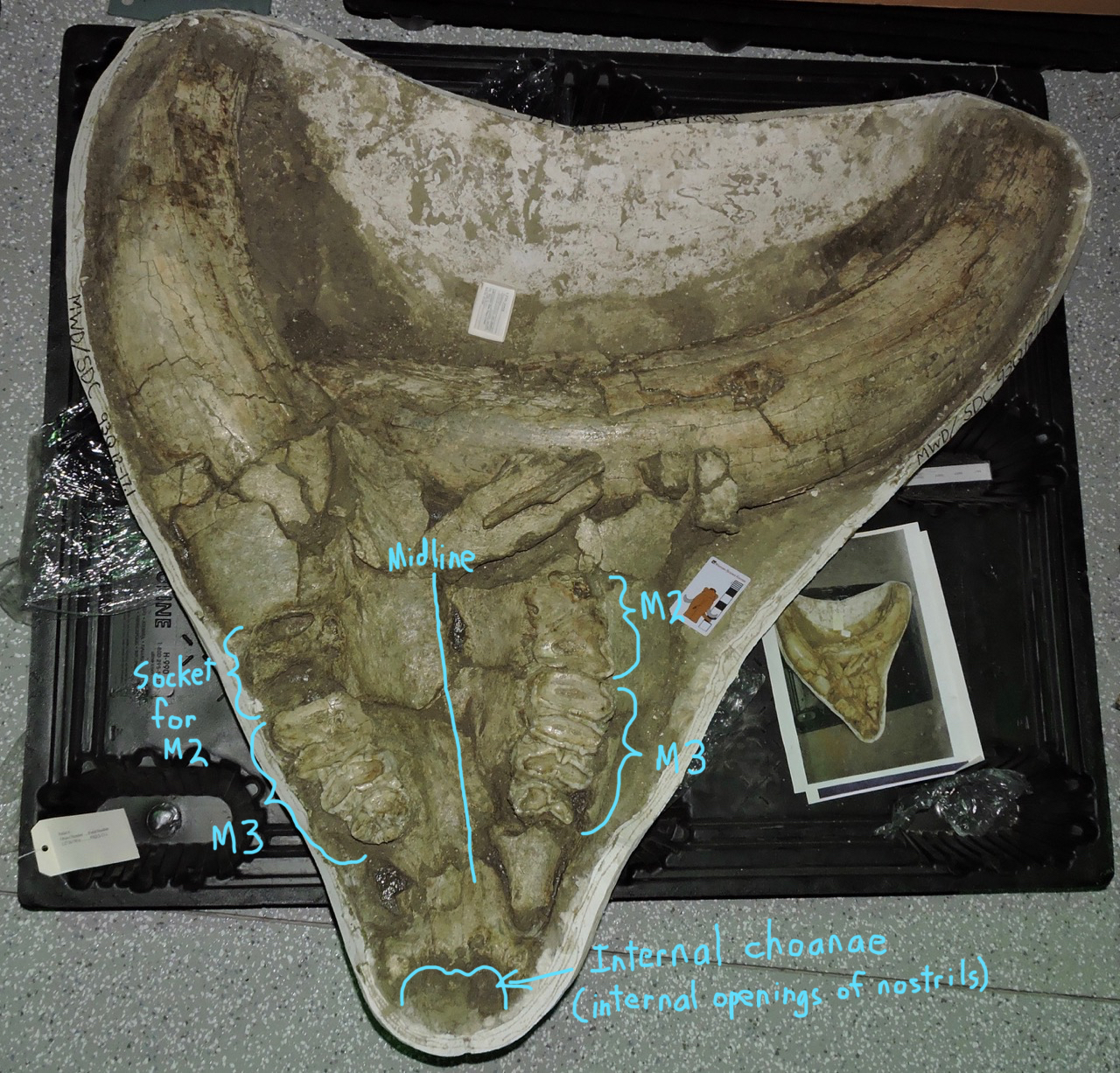 "Braces" was a fully mature mastodon, roughly 40 years old based on tooth wear. The right 2nd molar is missing, and I'm pretty sure it had fallen out well before the mastodon died. It looks like the socket had started closing up, and the corresponding left second molar was worn all the way down to the bone.Braces' name comes from anomalies in the wear pattern on his teeth. Typically, proboscidean teeth wear in a fairly predictable way. The upper teeth wear more rapidly along the interior half of the occlusal surface (the lingual, or tongue-ward, side), while lower teeth wear more rapidly along the exterior half (the labial, or lip-ward, side). This may seem a little confusing, but it just means that when the upper and lower teeth occlude they do so along an angled surface, as shown in this schematic drawing of a cross-section through a mouth:
 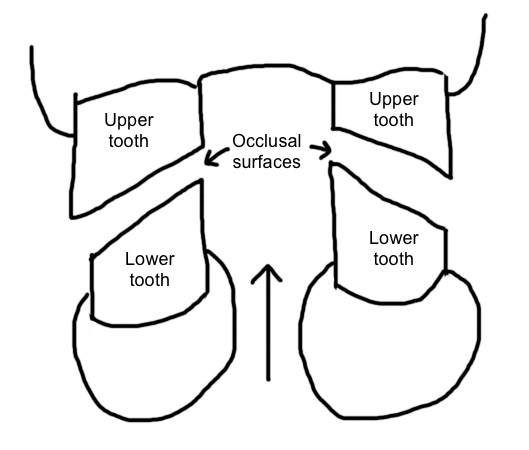 Here's a closeup of Braces' right 3rd molar, followed by an annotated version:
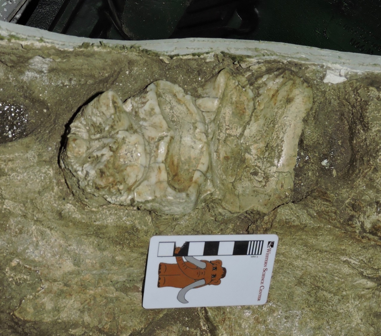
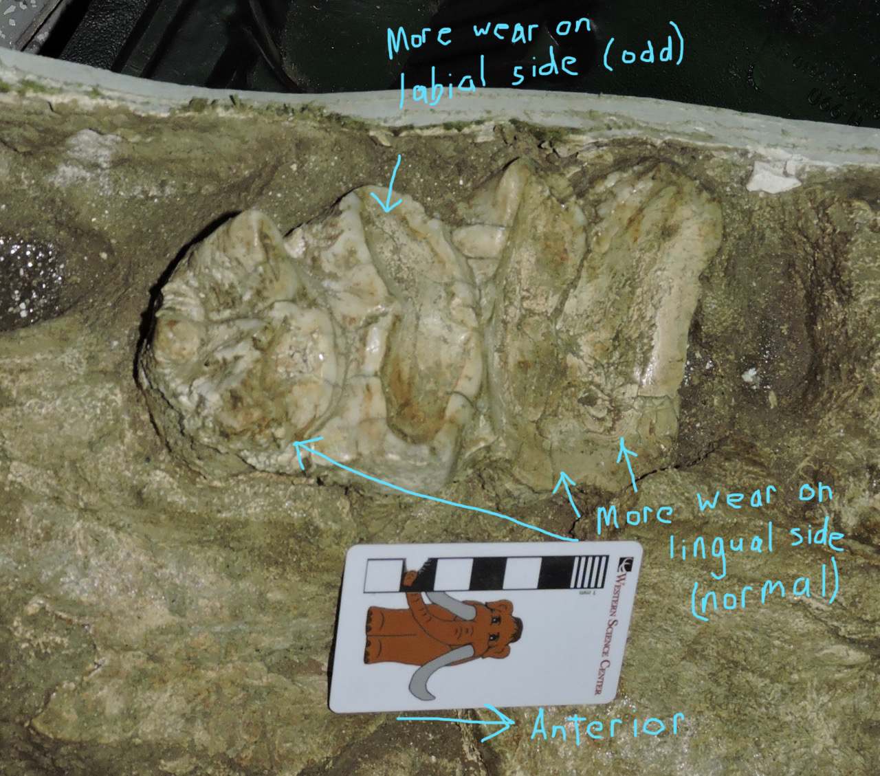 In Braces, the upper 3rd molar starts off wearing just as it should, with much heavier wear on the lingual side. But then, on the third loph, the wear pattern reverses, with heavier wear on the labial side like you would expect to see in a lower tooth. Then, on the fourth loph, the wear switches back to the normal pattern with heavier wear on the lingual side.It seems that there was some anomaly with the way Braces' teeth were aligned and occluding with each other. I'm not sure what caused this. Unfortunately we don't have the lower jaw, which might help. Maybe he had an injury to his lower jaw that changed the way he closed his mouth? Certainly Max, who was a slightly younger animal, had several jaw injuries. Or maybe this has to do with the right 2nd molar being lost before the left on; perhaps that sets up an asymmetry in the chewing dynamics. If this is the case, it's possible that if Braces had lived longer the pattern would have reversed itself. Maybe this is a relatively common but temporary condition, that only occurs during the brief periods when the tooth count is different on each side of the jaw.Along with most of our other mastodons, Braces will be on display in our Valley of the Mastodons exhibit that opens on August 5.
Fossil Friday - mastodon calcaneum
 Continuing with our mastodon spree in preparation for next month's "Valley of the Mastodons" workshop and exhibit, this week for Fossil Friday we have a mastodon calcaneum.This is a left calcaneum, seen above in posterior view. The calcaneum is the heel bone, and together with the astragalus makes up the ankle joint. In anterior view (actually anterior and slightly ventral, below) the two bright white surfaces are the articulations with the astragalus:
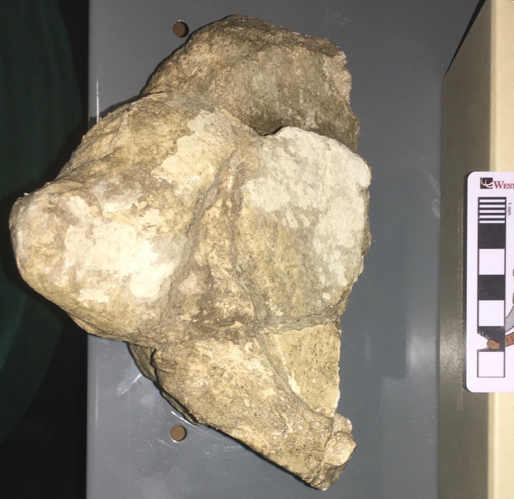 Here's a lateral view, with anterior to the left:
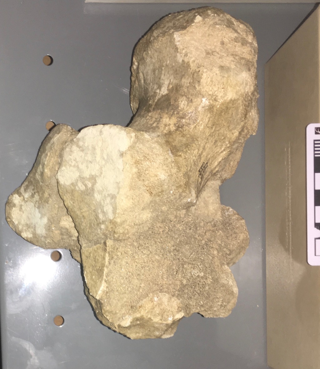 The large mass of bone sticking off the top is called the tuber calcis. Humans are plantigrade animals, meaning that we walk with our feet on the ground. In our feet, the tuber calcis projects down and back, forming our heel. We actually walk on the bottom of the tuber calcis (covered with a thin layer of soft tissue), while various muscles attach to the top surface of the tuber calcis through the Achilles tendon. When these muscles flex the Achilles tendon pulls the tuber calcis up, rotating the foot down. When a ballerina stands en pointe, she is flexing these muscles and keeping the tuber calcis rotated up and forward.Most large mammals are not plantigrade but are instead digitigrade; they are en pointe all the time. However, different limb geometry means that they don't have to constantly flex a group of muscles to stay in that position. Instead, the foot between the ankle and toes acts like an additional leg segment, moving forward and backward at the ankle and held vertically when at rest. (Some people mistakenly believe that animals such as deer and horses have "backward knees"; they're actually looking at the ankle joint, not the knee.)Surprisingly, for all their bulk, proboscideans are digitigrade. Even though the toes are short and stout, the ankle is held high and the calcaneum is off the ground. (See these fantastic CT-based videos of elephant foot bones from What's in John's Freezer to see the relationships of the bones to each other.) When you look at an elephant's feet, it may not immediately be clear that they're digitigrade because there is a huge fatty pad that covers the palm and heel, turning the whole foot into a column. In the hind foot, the top back part of the fat pad is nestled under the calcaneum.Even with their thick, columnar legs, elephants have a fair range of mobility at the ankle, and can move their feet forward and back relative to the shin. This is consistent with the large tuber calcis, which remember serves as a muscle attachment point for pulling the foot back (in this case, pulling it back to the vertical, resting position). What is a bit surprising is that, compared to elephants and mammoths, the tuber calcis in mastodons is enormous, roughly twice as large. Perhaps foot strength and dexterity was more significant in mastodons for some reason?This calcaneum, along with hundreds of other mastodon bones, will be on display in the "Valley of the Mastodons" exhibit at the Western Science Center next month.
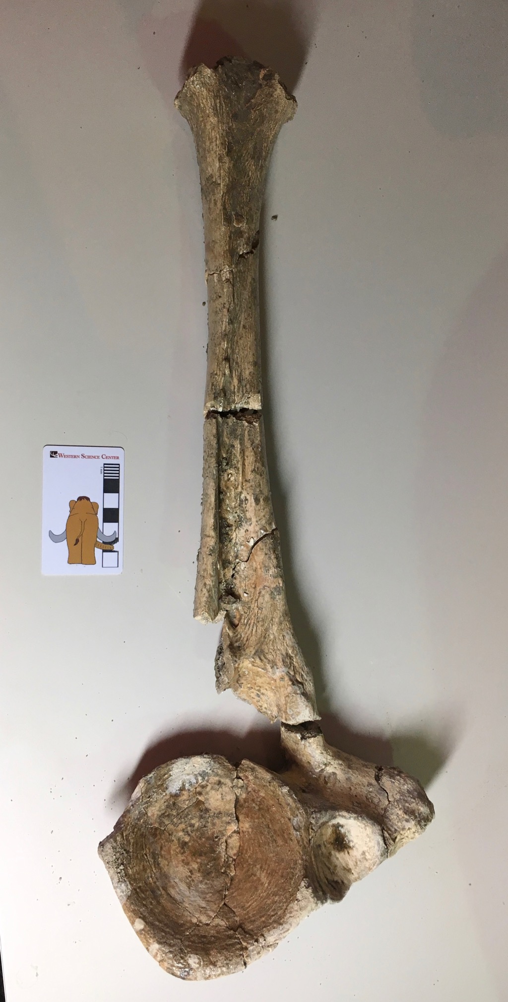
Fossil Friday - mastodon axis vertebra
 With the opening of the Valley of the Mastodons exhibit a little over a month away, we've started pulling specimens from the collections to work on layouts. The lab and collections workroom are full of mastodons!Today's specimen is a mastodon 2nd cervical (neck) vertebrae, more commonly referred to as the axis. It's shown above in anterior view, and below in posterior view:
 Animals with large heads face real biomechanical hurdles. If you're a quadruped, your head is stuck out on the end of a neck and is otherwise unsupported. This puts tremendous stress on the neck vertebrae, and requires massive muscles to hold up the weight, which affects the entire skeleton. In proboscideans the problem is even worse, because they have a large, heavy brain and, right out in front where it causes the most stress, massively heavy teeth, tusks, and trunk. Proboscideans have addressed this issue in two ways: the skull itself is actually pretty lightly built for its size, and the neck vertebrae are compressed front to back so that the neck is relatively short. But even with a short neck, elephants sometimes need to move their heads. So while the neck vertebrae are short, they have massive attachment areas for muscles and some range of mobility between the vertebrae. The huge, robust neural arch and spine on this mastodon vertebra reflect the large muscles attached to it. Just as a "gee, whiz!" point, the mass of bone that forms the top of the neural canal is roughly the same size as the entire centrum of a bison cervical vertebra, and it's not as if bison have tiny heads!In most mammals, nodding the head up and down largely involves movement between the skull and the first cervical vertebra (the atlas). In contrast, if an animal rotates its head along a longitudinal axis, such as the RCA dog below (from Wikipedia), or the elephant in this video, much of the movement is occurring between the atlas and axis vertebrae.
 (OK, the pedant in me has to point out that, while nodding and rotating largely involve the atlas and axis respectively, the rest of the cervical vertebrae are also involved in these movements, so injury or fusion at any point in the neck will cause a decrease in mobility. In addition, humans, because our bipedal stance has resulted in precariously-balanced and misshapen heads, work a bit differently. From a vertebral standpoint, our equivalent motion to what the RCA dog is would be looking from left-to-right, but when we cock our head to the side, that's the vertebral equivalent to looking left-to-right in other mammals. Finally, the necks in non-mammal tetrapods such as reptiles and birds work quite differently from mammals, so much of this doesn't apply to them.)The mastodon axis shown here is actually part of an associated skeleton that came from the West Dam of Diamond Valley Lake, which includes several other vertebrae, tusks, and a number of ribs. Like most of our mastodon material, it will be on exhibit starting in August.
Fossil Friday - mastodon caudal vertebra
 As promised, as we prepare for the Valley of the Mastodons workshop and exhibit, I'm going to be talking even more than usual about mastodons. We have hundreds of mastodon bones in the WSC collections, and we have to examine them all over the next month to decide what's going on exhibit (which will be most of them).The small bone shown above is a caudal (tail) vertebra, shown in dorsal view with anterior at the top. The ventral view is below:
 Looking at elephant tails for comparison (there isn't a lot of literature on mastodon tails!) this vertebra appears to come from roughly the middle of the tail. Proboscidean tails are relatively limited in use (modern elephants will use them to brush off insects, babies will sometimes hold onto their mother's tail, etc.), so the more distal vertebrae are quite simple. This vertebra has a neural canal (the passage for the spinal cord) that is open dorsally and very small transverse processes, both typical for proboscidean caudal vertebrae.In anterior view (below) we can see that the vertebra is also missing the epiphysis that forms the end of the bone:
 An unfused epiphysis is typically indicative of a bone that was still growing, suggesting that this was a relatively young animal. In fact, this vertebra is part of the associated skeleton of "Little Stevie", the most complete mastodon from Diamond Valley Lake. Based on his teeth and bone development, "Stevie" was a late adolescent, probably somewhere in the range of 18-22 years old.This vertebra, along with hundreds of other mastodon bones, will be on exhibit starting this August with the opening of Valley of the Mastodons.
Fossil Friday - mastodon molar
 This August we're opening a major new exhibit and hosting an associated workshop at Western Science Center, Valley of the Mastodons. In the lead-up to the exhibit/workshop, I'm going to be talking about mastodons even more than usual.The specimen shown above is a heavily-worn tooth in occlusal view. The anterior end of the tooth is at the top, and two corners are broken off. Based on the size, shape, and wear pattern, this appears to be an upper right 1st molar.Below is a lateral (labial) view, with anterior to the right:
 In this view it's clear that this tooth is almost completely worn away. The wear is heaviest at the anterior end, which is typical for proboscideans with horizontal tooth replacement. As this is an upper tooth, the wear is actually lighter on this side than on the medial (lingual) surface.This tooth was found along the San Diego Canal, apparently near the mastodon jaw we CT scanned a few weeks ago. However, it doesn't appear to be from the same animal, as that specimen had apparently lost its first molars some substantial amount of time prior to its death. This tooth, as well as nearly all of our other mastodon remains, will be on display this August in Valley of the Mastodons.
Fossil Friday - new mastodon CT scans
 Almost two years ago we took Max's lower jaw to California Imaging and Diagnostics for a CT scan. We got some fascinating data that raised a lot of new questions, so to help answer them we recently took a second mastodon to CID for scanning.The particular specimen is from the San Diego Canal, and includes a nearly complete lower jaw and a partial cranium. The cranium was of particular interest to us. It was broken in such a way as to include the entire left tooth row including the tusk, but the fragment was narrow enough to fit through the medical CT scanner; a complete adult mastodon skull is typically far too large to fit through the scanner. Based on tooth wear, while this mastodon was an adult he (it appears to be a male) was as much as 10 years younger than Max, so it gives us an interesting point for comparison. We're still examining the data, so what I'm posting here is preliminary (I only examined most of the data myself for the first time yesterday):Here's a transverse section through the SDC specimen's lower jaw, cutting through the last roots on the 3rd molars:
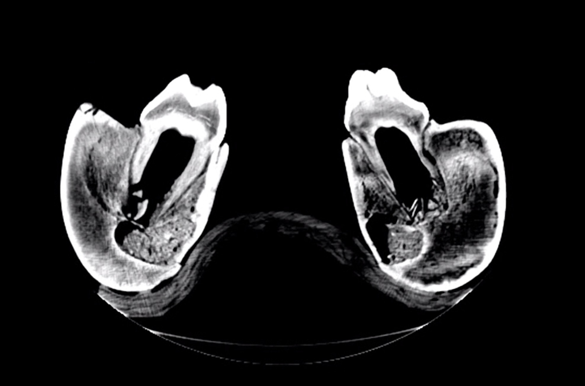 For comparison here's a similar section through Max's jaw:
 The SDC mastodon was a young animal, probably in his early 20's. His 3rd molars were still growing, and had large open pulp cavities and short roots. Max was a middle-aged mastodon in his mid-to-late 30's, with fully developed 3rd molars with deep roots and nearly closed pulp cavities.Here's a sagittal section through the SDC specimen's right dentary, followed by a similar section on Max:
 This view also emphasizes the huge open pulp cavity in the SDC specimen compared to Max's nearly solid teeth.Here's another sagittal section through the SDC right dentary, on a slightly different plane than the image above:
 Notice the differently-textured bone just in front of the 2nd molar, outlined in red below:
 With their horizontal tooth replacement, proboscidean teeth are constantly moving forward in the jaw, which means the bone in the jaw is constantly being absorbed and redeposited. The SDC specimen had recently lost his first molar, and the socket is filled by young, spongy bone that shows up as a different texture in the scan. In the much older Max this part of the jaw is as dense as the surrounding bone.Below is a transverse section through the cranium, followed by a marked-up version. I've rotated the image so that it's approximately in life orientation (it was upside down in the scanner):
 The blue area is the 3rd molar, at about the level of the 2nd lophid. The red area is actually the bottom of the unerupted part of the tusk, which extends all the way back at least to the 3rd molar. We can't tell in this specimen just how far back the tusk goes, because it's curving upward and the upper part of the cranium wasn't preserved.Finally, here's a section though a more distal part of the tusk:
 Again, this is rotated into approximately life orientation. One side of the tusk is damaged, and splinters of the tusk have broken off the inside and are laying haphazardly in the pulp cavity (which also contains some sediment). The better-preserved area shows visible concentric lines, the tusk's growth rings.We'll be studying these new scans in detail over the summer and fall, so there will be more to come. Thanks to CID for providing access to their CT scanner and running these scans for us!
