
 With over 100,000 Pleistocene fossils in the WSC collections, there are still many that I've never seen. There are others that I've glanced at dozens of times without ever taking a close look, like the mastodon vertebra shown here. As a result, interesting stories are often in plain sight, unnoticed until someone takes a close look.The bone is shown above in anterior view, and is a thoracic vertebra based on the rib articulation visible on the right side of the image. It appears to be a fairly posterior thoracic, although I haven't tried to identify the exact position. The vertebral centrum is heavily eroded, especially on the ventral side, but the dorsal part is better preserved. This vertebra was associated with a few scrappy remains of at least one other vertebra, but that was all that was recovered from this individual.In dorsal view we can see the base of the neural spine and the top of the neural canal. This is where things get a little strange:
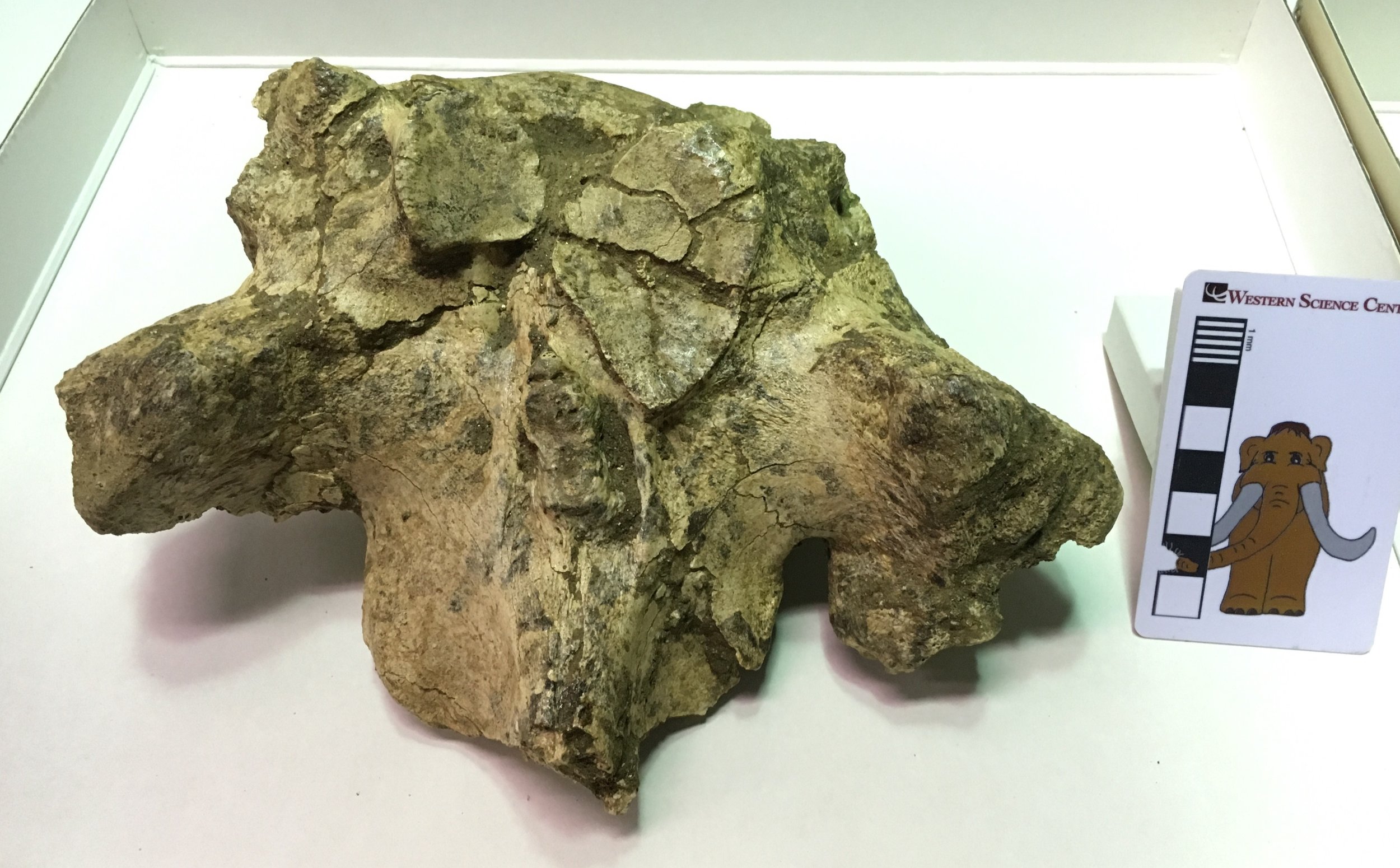 Typically, a vertebra articulates at three points with each vertebra in front of and behind it. Two of those points are the anterior and posterior surfaces of the centrum (so the anterior surface of the centrum articulates with the posterior surface of the centrum in front). The other articulations are located on the neural arch or spine. These articulations are often found at the tips of projections called zygapophyses, so the two prezygapophyses on the front edge of the neural arch will articulate with the corresponding pair of postzygapophyses on the next vertebra in front. The actual anterior articulation areas are called the prezygapophyseal articular facets (because zygapophysis was not an intimidating enough term by itself)!In mastodon thoracic vertebrae the prezygapophysis doesn't really stick out as a projection, but the articular facets themselves are very large. Below is the dorsal view image with the articular facets outlined in red:
 The left and right prezygapophyseal articular facets should be nearly mirror images of each other, but that's clearly not the case here. The right facet is more than twice as large as the left, and is actually so large that it extends slightly across the midline of the vertebra onto the left side. In addition, note the area outlined in blue, shown below in an oblique closeup:
 This is an osteophyte, a pathological bony growth that may form due to injury or as a result of osteoarthritis. It's not clear exactly what happened to this mastodon. The vertebra could have grown abnormally due to a genetic condition, and the resulting unusual stress caused by a deformed vertebra could have caused osteoarthritis later in life. Alternatively, the mastodon could have suffered an infection or injury that resulted in the deformed vertebra. The injury needn't have occurred in this spot; a muscle or bone injury elsewhere in the back or even in the legs or feet could have placed enough asymmetrical stress on the back to cause this deformation. Basically, walking with a limp over a long period of time could have potentially caused a condition like this. Since we have only a few bones from this animal, it's likely we'll never know for sure why this mastodon had such an asymmetrical vertebra.
