Fossil Friday

Fossil Friday - tusk fragment

IMGP1133.JPG
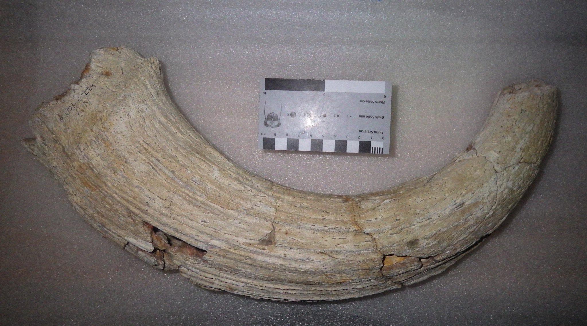
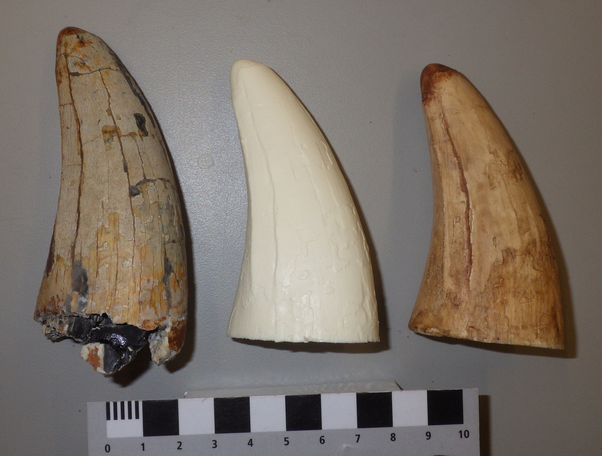
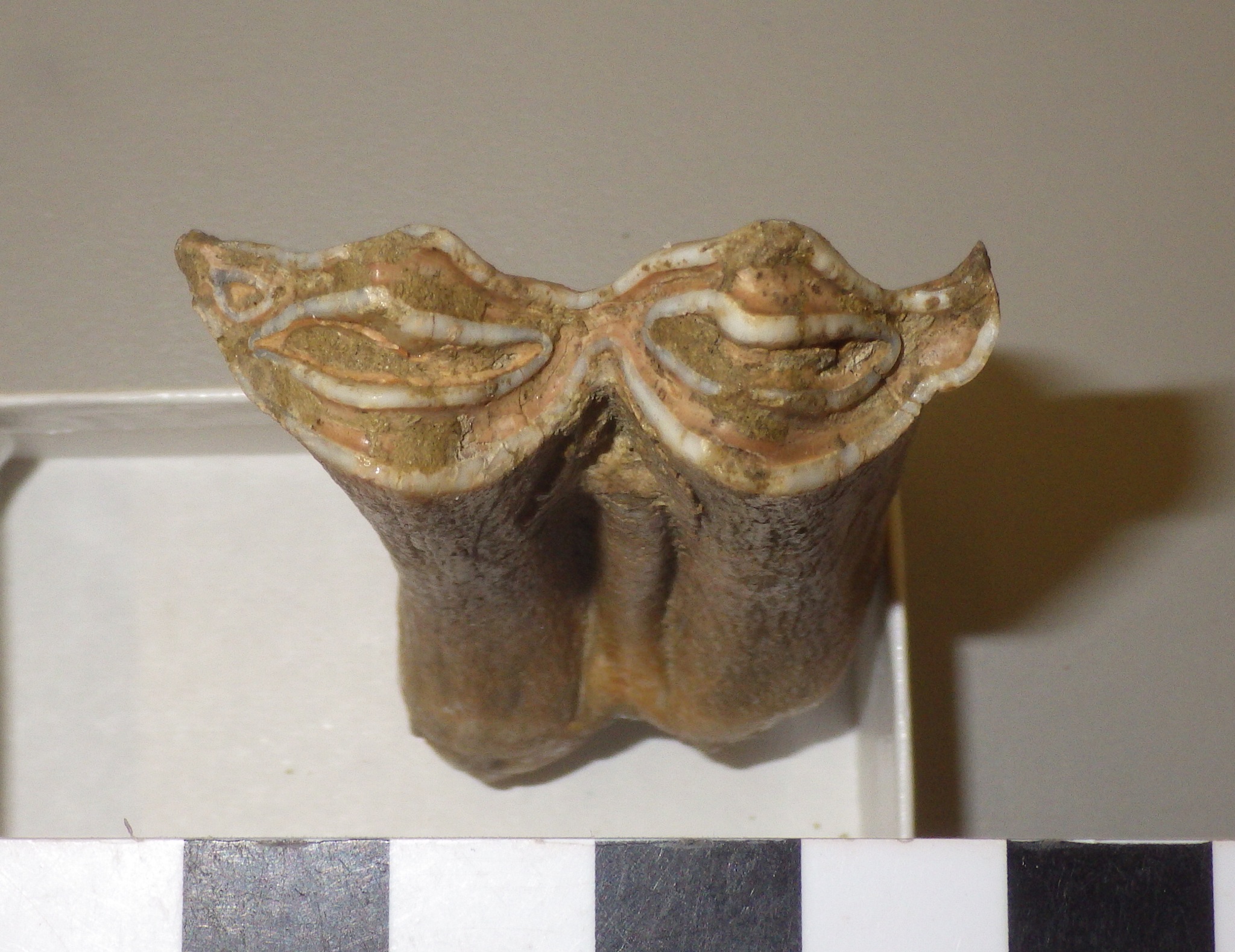
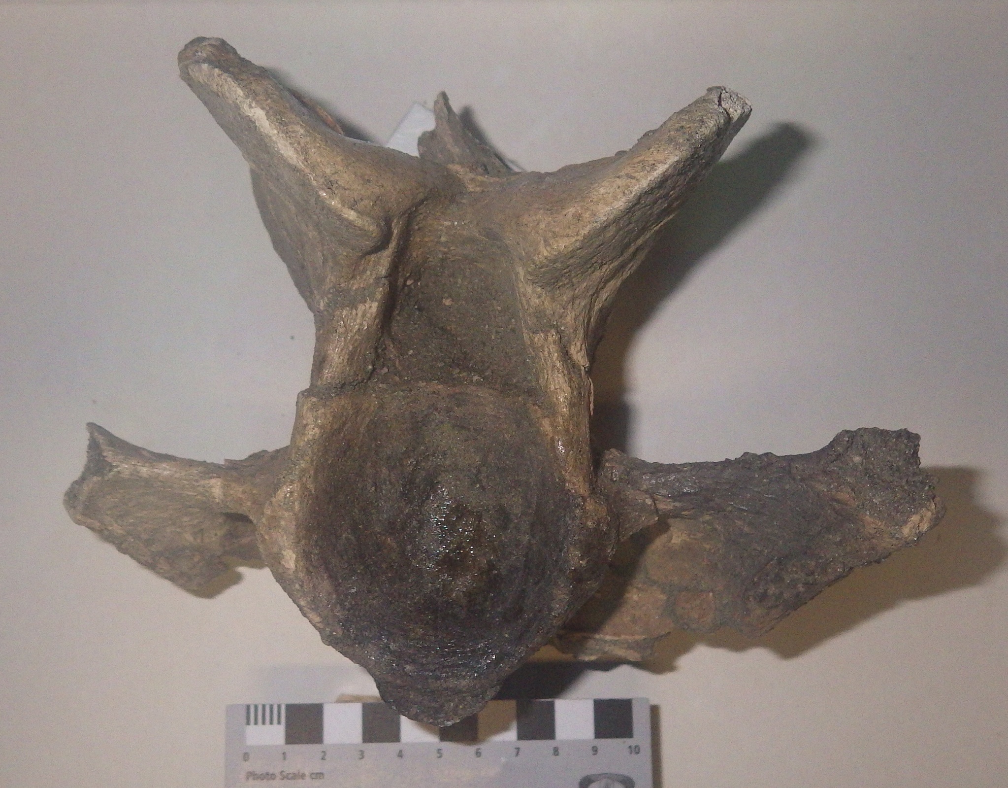
One of the prominent features of modern elephants are tusks, which are greatly enlarged upper incisor teeth. The presence of tusks is shared with most of the elephants' extinct proboscidean relatives, including mammoths and mastodons.Besides their size, elephant tusks are a bit different from most mammal teeth in that they lack enamel; tusks are made of ivory, which is dense dentine covered with cementum. Elephants use their tusks for a variety of tasks, including fighting, scraping bark off trees, and helping to loosen sediment when digging water holes, and the ivory tusks will get worn and polished at the tips. If the use is too vigorous the tip of the tusk may break off entirely. This isn't necessarily a problem for the elephant, because the tusks grow continuously. But it does mean that elephants may leave certain areas littered with broken tusk fragments.Mammoths and mastodons had tusks that were structurally very similar to those of modern elephants, and there they appear to have used them in similar ways. Their tusks were also just as prone to breakage, and isolated fragments often turn up as fossils. Shown above is the broken tip of a tusk that found near the western end of Diamond Valley Lake. Unfortunately, there are no morphological features present to tell if this came from a mammoth or a mastodon (at least none that I can identify). The tip (on the left) is polished and rounded from heavy wear, while the broken edge on the right is sharp and jagged. Below is a view of the other side of the same fragment:

IMGP1134.JPG
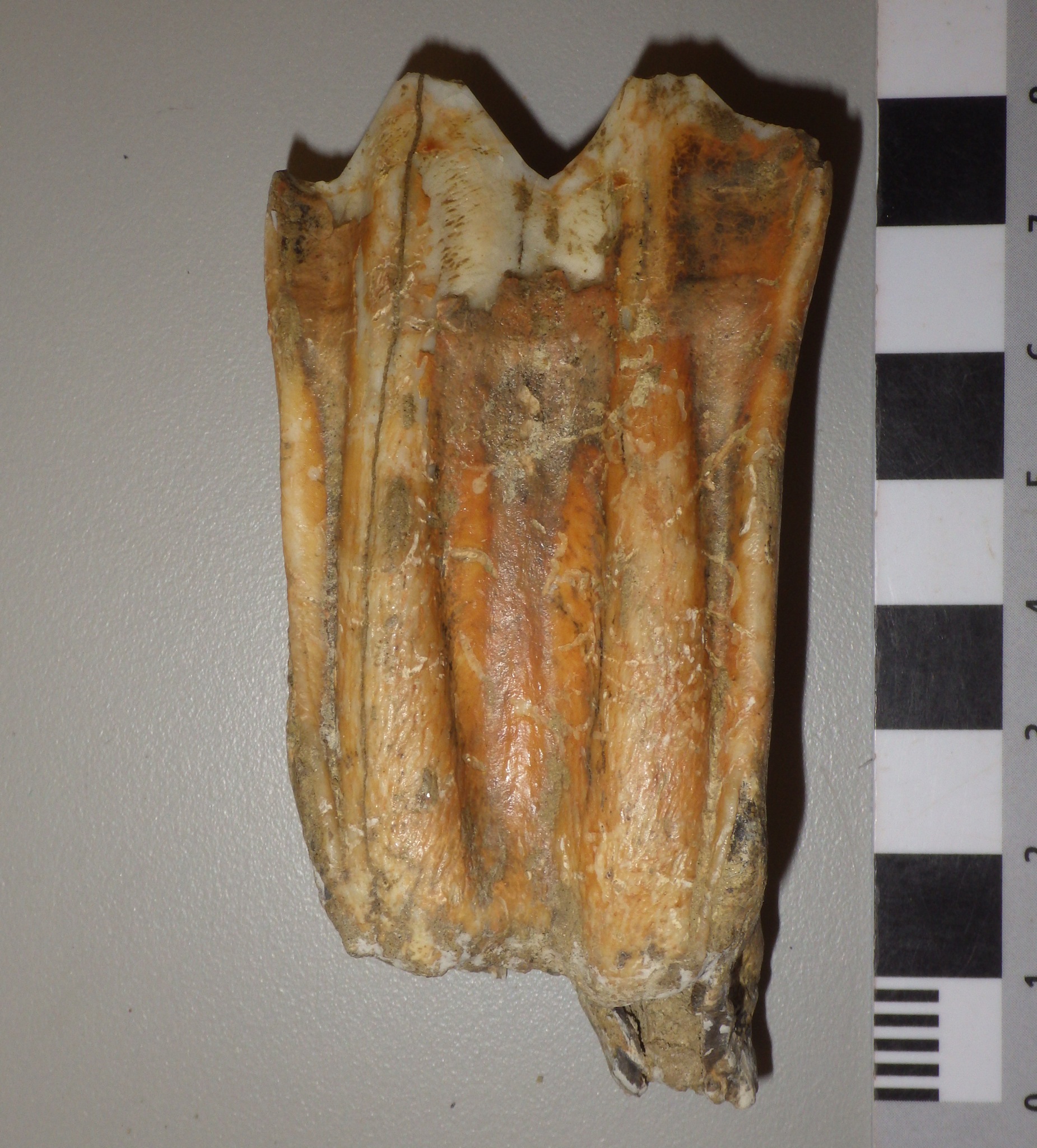
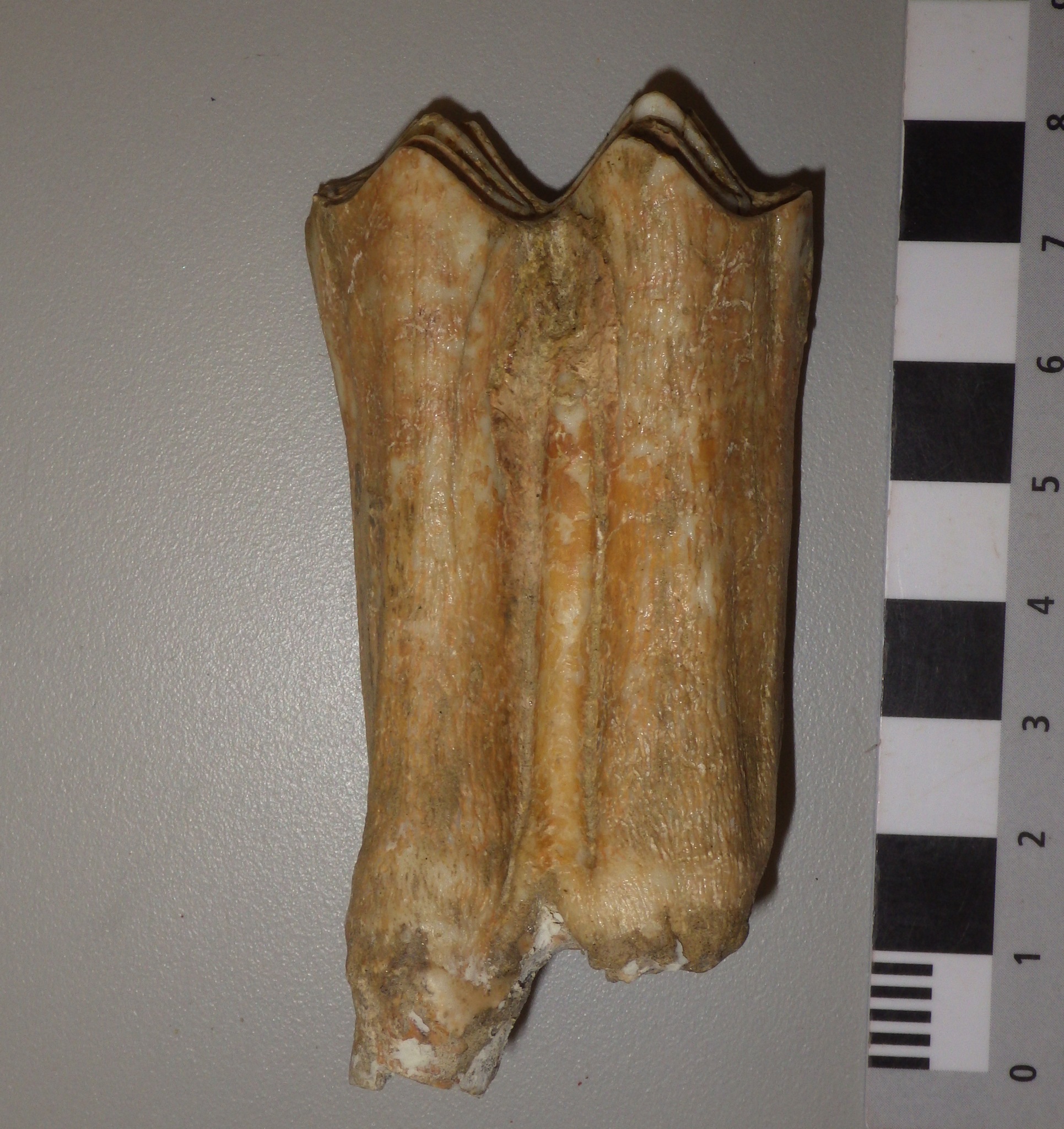
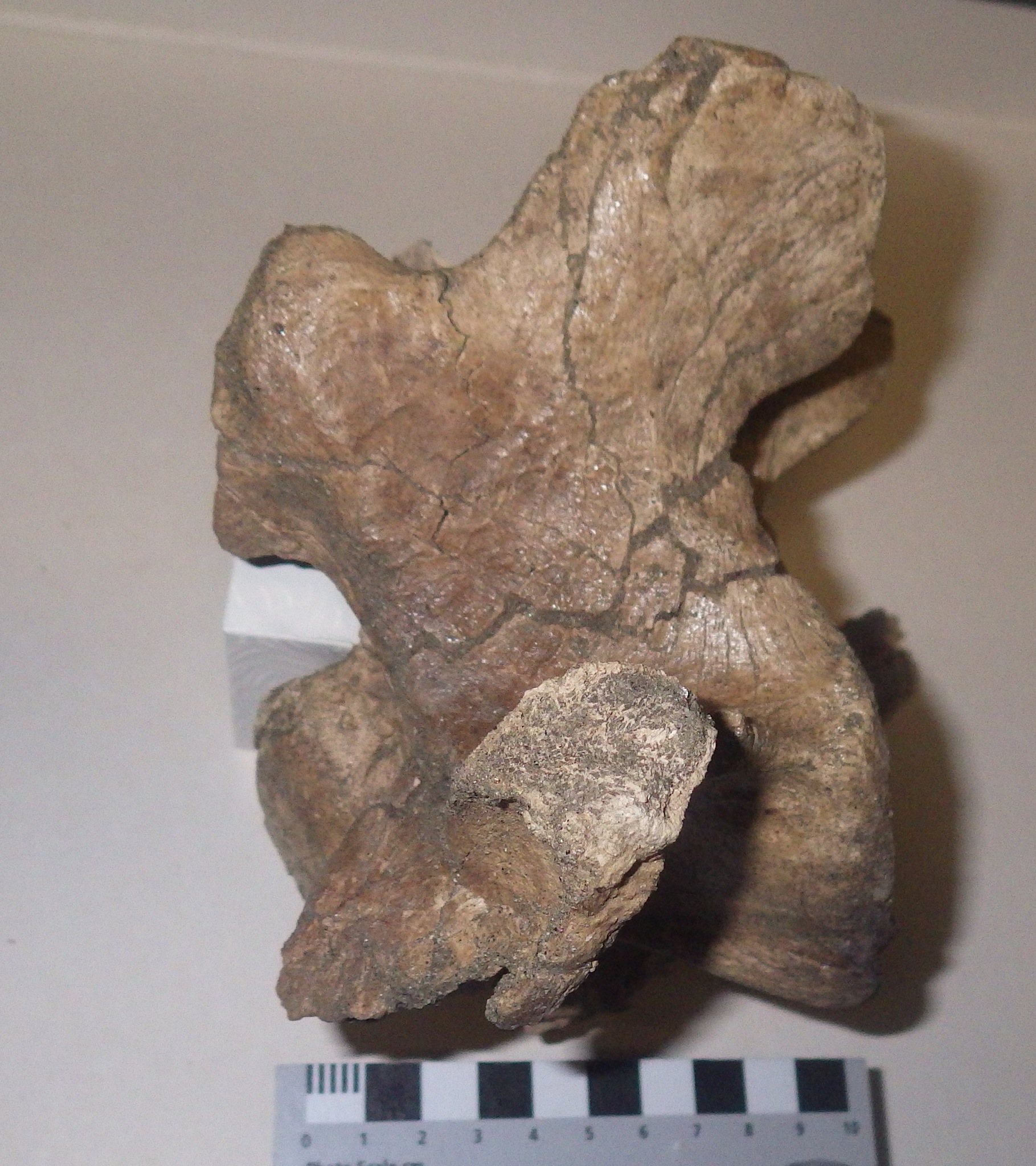
This surface has a wrinkled appearance, and a rounded groove running for most of its length. I believe this represents an earlier break, in which the broken surface was smoothed off and polished prior to the entire tip breaking off. If that's correct, then this small fragment actually records two tusk-breakage events with a substantial amount of time between them.It's interesting to note that, according to Haynes (1991), while African elephants use their tusks for a variety of activities, they normally only break them when fighting each other. This seems to be especially prevalent drought years, when elephants will fight each other over access to water holes, although male elephants will fight in dominance displays, and there's evidence that mammoths and mastodons engaged in similar activities.Reference: Haynes, G., 1991. Mammoths, Mastodonts, and Elephants. Cambridge University Press, Cambridge, 413 p.

Fossil Friday - bison horn

IMGP1129.JPGBison are among the most common animals in the Diamond Valley Lake Local Fauna. While no complete bison skeletons were recovered during excavations for the construction of the reservoir, large numbers of fragments, individual bones and teeth, and associated bones were discovered. Some of these are impressively large, including the horn core shown here.Bison are members of the Family Bovidae, a diverse group that includes cattle, goats, sheep, and antelope. A characteristic shared by practically all bovids is the presence of horns in males (there are some domestic bovid breeds in which the males lack horns); in many species the females have horns as well. The horns vary greatly in size and shape between species, but they always have certain features in common: they form as outgrowths of the frontal bones on the roof of the skull, they do not branch, and they are covered in a keratin sheath. Keratin doesn't usually preserve in fossils, so what we generally see in fossils is the rough bone underneath. Because the horn is part of the frontal bone, it is not shed each year like the antlers of deer.There are two different extinct species of bison known from Diamond Valley, Bison antiquus and Bison latifrons; among other differences, B. latifrons is larger and has much longer horns. As big as this specimen is, it's not immediately clear which species it represents. It's on the small side for B. latifrons, but a little larger than some of our B. antiquus specimens.

Fossil Friday - mastodon premolar

IMG_0884.JPGI'm starting 2015 at Valley of the Mastodon with an example of the blog's namesake, a tooth from a mastodon.This tooth is much smaller than the one I showed for Fossil Friday back in October, and represents a much younger animal. Like elephants, mastodons grow a total of six teeth in each quarter of their jaws (not including tusks), deciduous premolars 2-4 and molars 1-3. However, these teeth erupt sequentially as if they were on a conveyer belt, so there are never more than three teeth in the mouth at any one time.The tooth is shown above in occlusal view, with the front of the tooth to the right. It seems to have been rolled around in a stream or otherwise eroded, as there is a fair amount of damage including missing roots. There is also a lot of wear on the tooth caused by chewing, so that the enamel ridges characteristic of mastodons have been worn down, leaving three oval enamel ridges on the occlusal surface of the tooth. The fact that there are three of these enamel ovals suggests that this was not the 2nd or 3rd premolar, as these teeth usually only have two enamel ridges. The tooth is also small, only about 7 cm long, and is about twice as long as it is wide, suggesting that it is the 4th premolar. Moreover, the enamel ridges are not perpendicular to the long axis of the tooth, suggesting that this is a lower premolar rather than an upper one.Below is a labial view of the same tooth, showing the heavily worn crown and the damaged roots:

IMG_0885.JPGBecause of the sequential tooth replacement in mastodons and elephants, the 4th premolar is only present in the mouth for a fairly short period of time. For modern elephants this tooth is only present from about 3 to 10 years of age. Assuming mastodonts grew at the same rate as elephants, this tooth probably represents a mastodon that was roughly 10 years old.

Fossil Friday - Tyrannosaurus tooth

IMGP1058.JPGThe vast majority of the Western Science Center's fossil collection comes from Pleistocene deposits in Riverside County, so most of our specimens are less than 200,000 years old. Being so geologically young, birds are almost the only dinosaurs in our collection, as birds were the only dinosaurs to survive the mass extinction at the end of the Cretaceous Period 65 million years ago. We do, however, have a handful of much older, non-avian dinosaurs at the museum.On the left above is a partial single tooth from Tyrannosaurus rex from the Late Cretaceous Hell Creek Formation in Montana, which was collected by Harley Garbani and donated to the museum by Harley and his wife Mary. The tooth was apparently found isolated, which is not uncommon with dinosaurs as they shed teeth throughout their lives. As is typical of Tyrannosaurus and other theropods, the tooth has serrated cutting edges, one of which is visible near the left edge of the tooth. There is noticeable apical wear at the tip of the tooth, which is somewhat rounded off.This tooth was one of the first subjects for our molding and casting program. At the center of the photo is an unpainted resin cast of the tooth, and on the right is a painted cast. The serrated cutting edge is actually more visible in the cast than in the original specimen.The original specimen is on display at the Western Science Center as part of the "Harley Garbani: Dinosaur Hunter" exhibit. Replicas of the tooth will be available in the museum store early next year.

Fossil Friday - bison molar

IMGP1053.JPGAs our molding and casting program gets underway, we've been going through the WSC collections looking for good candidates for molding. We've been particularly interested in teeth, because in many mammals the teeth are highly distinctive and potentially provide a lot of information about the animal. Over the last week we've been focusing on bison teeth.Shown above is a more-or-less complete lower right molar from Bison, probably the 2nd molar, shown in labial view. Here's the same tooth in lingual view:

IMGP1052.JPGIn this view there's a ridge running vertically down the middle of the tooth called a stylid. This is a column of enamel that will show up on the occlusal surface of the tooth as a small ring of enamel if the tooth is sufficiently worn. This stylid seems to always be present in Bison, but in the closely related genus Bos (cows) the stylid is usually absent or only weakly developed.Here's the tooth in occlusal view:

IMGP1055.JPGBison have a selenodont tooth pattern with two main crescent-shaped cusps, similar to many other artiodactyls. The tooth is worn, but only slightly, so this animal was likely a young adult when it died. The tooth had not yet worn down as far as the stylid on the lingual surface, so the enamel loop isn't visible; had the animal lived longer the loop would eventually have appeared as the tooth wore down.This tooth was found near the east dam of Diamond Valley Lake.

Fossil Friday - peccary molar

IMG_0879.JPGThe Pleistocene megafauna from Diamond Valley Lake is dominated by a few prominent groups such as horses, bison, and mastodons. But there were a lot of species of mammals in the valley that are represented by only a limited number of fossil specimens. One of these rare types is the peccary, Platygonus.Peccaries, or javelinas, are artiodactyls that are sometimes referred to as "New World pigs". In fact, they are placed in their own family, the Tayassuidae, separate from domestic pigs and other Old World pigs (Family Suidae). Peccaries still live in Central and South America, and one species, the collared peccary, can still be found in the southwestern United States (the example below is at the Henry Doorly Zoo in Omaha):

IMG_0880.JPGPeccaries were much more diverse and widespread in North America during the Pleistocene, and several different genera are known from the fossil record. Various species of the extinct genus Platygonus have been found all over the United States (the skeleton below was on display at the Smithsonian Institution):

IMG_0877.JPGThe Platygonus tooth shown at the top is an upper molar, probably the upper right first molar. It's only lightly worn, as is clear in lateral view:

IMG_0882.JPGThis tooth was found near the east dam of Diamond Valley Lake, not far from where the museum is located.

Fossil Friday - horse toe

IMG_0868.JPGSome of the most common animals in the Pleistocene deposits around Diamond Valley Lake are horses. While we don't have any complete individual horse skeleton in the WSC collections, across all our different specimens we probably have just about every bone in the skeleton represented. Shown here is an isolated horse hoof.Horses have highly specialized feet, with only a single toe on each foot. That toe is Digit III, equivalent to our middle fingers (on the front feet) and our middle toes (on the back feet). Modern horses actually also have non-functional remnants of the 2nd and 4th toes, a relict of their multi-toed ancestors.Fingers and toes are made up of a series of bones called phalanges (singular: phalanx) that articulate end-to-end with each other; the articulations are the joints in our fingers and toes. In horses, the last phalanx, or ungal, is enlarged into an arc-shaped wedge of bone to support the weight of the animal. In life, the ungal is covered with a keratinized sheath, the hoof itself, which doesn't usually preserve in fossils.The image at the top shows a horse ungal in dorsal view, with anterior at the top. The scalloped area at the back of the bone is the articulation with the next phalanx in the toe.Here' same ventral view of the same bone:

IMG_0867.JPGThe lateral view, showing the wedge shape (the articulation with the next phalanx is at the upper right):

IMG_0865.JPGNote the how symmetrical this bone is in dorsal and ventral view. Horses are the only hoofed mammals in the valley that have a single toe, and as such are the only ones with a more-or-less symmetrical hoof-shaped ungal. Other hoofed mammals from these deposits, such as bison, have two toes with much more asymmetrical ungals.

Fossil Friday - turkey

IMGP1010.JPGIt's the day after Thanksgiving in the US, so for today's Fossil Friday we have a probable turkey bone from south of Diamond Valley Lake.I found this bone by accident last week while looking for good examples of packrat teeth. While this fragment isn't much to look at, we have almost no other fossil turkeys in the WSC collection.This fragment is part of the distal end of the tarsometatarsus. This is a structure unique to birds and certain other dinosaurs, in which the ankle bones (the tarsals) and the foot bones (the metatarsals) are fused into a single unit. The proximal end articulates with the shin bone (tibiotarsus in birds), while the distal end has structures called trochlea that articulate with the toes. Most birds have three toes, and so have three trochlea at the end of the tarsometatarsus, as can be seen in the examples from modern turkeys shown below (from Thornton et al. 2012):

IMG_0864.JPGThe WSC fragment is one of the trochlea, I think for either the second or third digit (the inside or middle toe). I'll need to compare it to a modern turkey bone to confirm which one, and unfortunately the turkeys you buy at the grocery store don't typically include the tarsometatarsus!Judging by their near-absence in our collection, it would seem that turkeys were relatively rare in the valley during the Pleistocene. This is a little surprising since vast numbers of turkey bones have been recovered from Rancho la Brea, which is only about 80 miles away from Hemet (more than 11,000 turkey bones have been found there, according to Bocheński and Campbell, 2006). The Rancho la Brea turkey was long considered to be its own genus, Parapava, but was reassigned to the same genus as the extant turkey as Meleagris californica by Steadman (1980). (Below is a reconstructed skeleton of M. californica on display at the George C. Page Museum.)

IMG_0863.JPGIt's possible that turkeys may have breeding at Rancho la Brea, and there are reasons to think that they may have been particularly susceptible to being trapped in tar (Bocheński and Campbell, 2006). It's also not clear that the turkey in the WSC collection is M. californica; it could be the modern wild turkey M. gallopavo or some other species. Even so, it's remarkable that turkeys were seemingly quite rare such a short distance away from Rancho la Brea, and may have interesting implications for different environmental conditions across California during the Pleistocene.References:Bocheński, Z. M. and K. E. Campbell, Jr., 2006. The extinct California turkey, Meleagris californica, from Rancho la Brea: Comparative osteology and systematics. Contributions in Science, Natural History Museum of Los Angeles County, No. 509:1-92.Steadman, D. W., 1980. A review of the osteology and paleontology of turkeys (Aves: Meleagridinae). Contributions in Science, Natural History Museum of Los Angeles County, No. 330:131-207.Thornton, E. K., K. F. Emery, D. W. Steadman, C. Speller, R. Matheny, and D. Yang, 2012. Earliest Mexican Turkeys (Meleagris gallopavo) in the Maya Region: Implications for Pre-Hispanic Animal Trade and the Timing of Turkey Domestication. PLoS ONE 7(8): e42630. doi:10.1371/journal.pone.0042630

Fossil Friday - packrat molar

IMGP1014.JPGFor this week's Fossil Friday I'm sticking with teeth. In contrast to last week's large camel molar, this time I chose the other end of the size spectrum with a tiny tooth that's only about 3 mm long.For all the attention that large animals receive, rodents and other small mammals are probably the most common vertebrate fossils in the Pleistocene deposits around Diamond Valley Lake. One of the more common rodents in our collection is the genus Neotoma, the packrat.Packrats (or woodrats) are in the Family Cricetidae, the same family that includes voles, lemmings, and hamsters. They shouldn't be confused with the brown rat (genus Rattus), which is what many people think of when they hear "rat". Rattus (including the white forms commonly used in laboratories) is a member of the Family Muridae. Rattus is only distantly related to the cricetids and was introduced into the Americas by humans.Packrats have a reduced dentition, with only one incisor and three molars in each half of both the upper and lower jaws. The molars have five cusps that are arranged in such a way that as they wear down they form three enamel-edged oval basins in occlusal view:

IMG_0860.JPGDifferent genera have distinctive cusp patterns, and in fact each individual tooth position has a unique, easily-identified pattern (see the images at this site). This particular tooth is the first lower right molar from Neotoma.There are a number of species of Neotoma known from California, and unfortunately the different Neotoma species are not as easily identified on the basis of an individual tooth. So, at least for now, this tooth can only be identified as Neotoma sp.

Fossil Friday - camel molar

IMGP0983-4.JPGFor this week's Fossil Friday we'll return to camels, specifically the large extinct camel Camelops hesternus that's pretty common in Pleistocene deposits in California.This specimen is an upper molar collected near the west dam of Diamond Valley Lake. I'm pretty sure this is the upper right first molar, although I can't yet rule out the possibility that it's the second molar. This tooth was found associated with several other molars and premolars, as well as small skull fragments that all appear to be from one individual.The image above is in occlusal view, showing the chewing surface. The tooth is fairly heavily worn, showing the pattern of folded enamel ridges; the shiny grayish-white ridges are enamel, with softer dentine in between. By having these alternating areas of hard enamel and softer dentine, the enamel ridges always stick out slightly beyond the occlusal surface, so the tooth maintains a sharp chewing surface even as it's being worn down. The elevated enamel ridges are especially noticeable in lingual view (since this is an upper tooth, the occlusal surface is at the bottom):

IMGP0985-0.JPGThe particular pattern of enamel ridges is one of the primary means of distinguishing between different groups of mammals, even down to individual species. For some species it's possible to make an identification on the basis of a single well-preserved tooth.Camels have a general tooth pattern that's called selenodont, in reference to the crescent-shaped enamel ridges in occlusal view. Camels share the selenodont pattern with many of the camel's artiodactyl relatives, including cervids (deer) and bovids (bison, cows, antelopes, etc.).Just for the sake of completeness, here's the labial view of the same tooth:

IMGP0984-0.JPG

Fossil Friday – bison cervical vertebra

IMGP0966.JPGFor today's Fossil Friday we have a vertebra from one of the more common Pleistocene animals in the region, the bison.The vertebral column (backbone) in mammals is typically divided into five separate regions, based on common characteristics and the location in the body. From front to back these are the cervical vertebrae, which form the neck; thoracic vertebrae, which make up part of the back and have ribs attached; the lumbar vertebrae, which form the lower back; sacral vertebrae, which form part of the hip girdle; and caudal vertebrae, which make up the tail.Nearly all mammals have vertebrae from each of these regions. The main exceptions are whales and sea cows, which have no sacral vertebrae. (In fact, they almost certainly do have sacrals, but they are modified to the point that they're indistinguishable from lumbar vertebrae. Very primitive whales and sea cows still had identifiable sacrals.) Even tailless mammals such as humans still have caudal vertebrae; our tailbone, or coccyx, is formed by five fused caudal vertebrae.Another peculiarity of mammals is that almost every living species has exactly 7 cervical vertebrae, whether the neck is short, long, rigid, or flexible. In many animals the cervical vertebrae are very intricate, with all kinds of slender projections and holes passing through them. This happens because a lot of things are happening in the neck. Besides the spinal cord passing through the vertebrae (as it does in all vertebrae) the carotid arteries and jugular veins run alongside the cervicals as they carry blood to and from the brain, both the esophagus and trachea pass directly under the cervicals, and there are numerous muscles and their associated nerves and tendons that control the complex movement of the head and neck. With so much going on, these vertebrae have to be complex to make room for everything while still providing attachment areas for all the neck muscles.The image at the top is an anterior (or cranial) view of a bison cervical vertebra. The bone should be close to symmetrical, but has been somewhat deformed, probably after burial. The prominent oval structure near the bottom is the anterior articulation, the ball part of a "ball-and-socket" joint with the next vertebra forward. Immediately above that is a dark circular area (dark because it's still filled with sediment) called the neural canal, which is the passage for the spinal cord.Here's a posterior (caudal) view of the same vertebra:

IMGP0967.JPGThe round area at the bottom is the posterior articulation, which is a concave socket that articulates with the ball from the next posterior vertebra. The sediment-filled neural canal is the bell-shaped structure immediately above the articulation.This is the left lateral view, with anterior to the left:

IMGP0968.JPGThe vertebra should actually be quite a bit taller, but most of the neural spine is missing. The neural spine is a prominent bony projection along the top of the vertebrae that serves as an attachment point for some of the muscles responsible for raising the head.The label on this specimen identifies it as the fourth cervical vertebra, which looks like a pretty good match to me (compare with the modern bison vertebrae shown here). There are two different species of bison known from Southern California, the long-horned Bison latifrons and the (relatively) short-horned Bison antiquus. Based on its size this vertebra probably came from the smaller B. antiquus.This vertebra was collected close to the same locality as the mastodon tooth from last week's Fossil Friday, about 15 miles southwest of the museum.

Fossil Friday - mastodon lower molar

IMGP0894.JPGFor this week's Fossil Friday we have a tooth from this blog's namesake, the mastodon Mammut americanum. This particular tooth was collected about 15 miles southwest of the museum, not far from Temecula in Riverside County.This tooth is the lower left 3rd molar. The image above is the side, or lateral view, often referred to in teeth as the labial view (literally "near the lips"). The front of the tooth is to the left, and a lot of wear is visible on the crown at the anterior edge. Below is the same tooth in medial, or lingual view (literally, "near the tongue"):

IMGP0896.JPGIn this view the front of the tooth is to the right, and the wear is much less obvious. The discrepancy in wear between the lingual and labial sides of the tooth are even more obvious when looking at the chewing surface of the tooth (occlusal view):

IMGP0895.JPGThe front of the tooth is to the left. Mastodon teeth have enamel cusps arranged into a series of transverse ridges. There are five of them on this tooth. The first ridge is almost completely worn away, especially on the labial side where the enamel is completely gone. The second ridge is also heavily worn. The third and fourth ridges show moderate wear, with only the top of the enamel ridges worn away, while the small fifth ridge has very little wear. In occlusal view an unworn mastodon tooth is completely covered in enamel, but as the tooth is used the enamel wears away, exposing the softer underlying dentine. In the image below the dentine is colored red, giving an idea of how much enamel has been lost:

IMG_0828.JPGThere are a few things that we can say about this mastodon based on the variations in wear patterns across this tooth. Because of the way mastodon teeth occlude when the mouth is closed, it's not unusual for the labial side of the lower teeth to wear more rapidly that the lingual side. The upper teeth usually show the reverse, with heavier wear initially on the lingual side. In both the upper and lower jaws the teeth eventually wear down until are essentially flat, but his tooth never reached that level of wear.The difference in wear across the tooth from the front to back is a result of the unusual method of tooth replacement in elephants and many other proboscideans, including mammoths and mastodons. Instead of having a mouthful of teeth all at once, mastodons grow in their teeth gradually and sequentially. As a tooth wears down, it moves forward in the jaw, eventually falling out and being replaced by the next tooth in the sequence. Since an individual tooth moves into position gradually, the front part of the tooth starts wearing down before the back part of the tooth has erupted. That's what we see in this tooth, in which the first enamel ridge is almost completely worn away while the last enamel ridge has almost no wear at all.Because of the sequential tooth replacement, we can also say a little about the age of this mastodon when it died. This tooth is the 3rd molar, which is the last one in the sequence, and it shows only a moderate amount of wear. This suggests that this mastodon was a fully mature adult, but not elderly, when it died. In modern elephants the 3rd molar doesn't erupt and begin to wear until the animal is about 25-30 years old. Assuming mastodon growth and tooth replacement rates were comparable to living elephants, this mastodon would likely have been 30-40 years old when it died.

Fossil Friday - Cretaceous invertebrate donation

IMGP0885.JPGLast Wednesday, Hemet resident Jeanette Hughes visited the Western Science Center to donate a box of Cretaceous invertebrate fossils that she and her late husband Richard collected more than a decade ago in Texas.Most of the specimens in the donation were ammonites, extinct cephalopods related to the modern chambered nautilus:

IMGP0887.JPG

IMGP0884.JPGThere were also several sea biscuits, a type of echinoid related to sand dollars:

IMGP0889.JPGA number of species of bivalve mollusks were represented, including oysters from the genus Gryphaea (or a close relative):

IMGP0888.JPGOne of the oysters is impressively large (note the 10 cm scale bar):

IMGP0890.JPGJeanette was able to provide us with pretty precise locality information for these specimens, making them potentially much more useful from a scientific standpoint. WSC did not have much of material from these taxa or from this time period, so this is a nice addition to our collection. I'd like to thank Jeanette for her kind donation.

Fossil Friday - Harlan's ground sloth jaw

IMGP0873.JPGFor this week's Fossil Friday we have a partial lower jaw of Harlan's ground sloth, Paramylodon harlani, collected near the eastern end of Diamond Valley not far from the museum's current location.This particular fragment is the back half of the left dentary (the left and right dentaries are the bones that make up the lower jaw in mammals). The image above is the lateral (side) view with the front to the left. The small projection sticking out on the middle of the back edge is the mandibular condyle, the part of the jaw that forms the joint with the rest of the skull.Here's the medial view of the same jaw:

IMGP0872.JPGAnd here's the dorsal (top) view:

IMGP0875.JPGIn this view we can see that, while the teeth are missing, the tooth sockets for the 3rd and 4th molars are preserved, as well as the back and inner edges of the socket for the 2nd molar. The mandibular condyle also appears much larger from this angle, because while it is relatively short vertically it is quite wide transversely (from side-to-side).There are several different species of sloths known from the Diamond Valley Lake region and that are represented in the WSC collections. Paramylodon harlani is by far the most common, making up over 93% of the individual sloth bones from the valley (Springer et al. 2010).Reference:Springer, K., E. Scott, J. C. Sagebiel, and L. K. Murray, 2010. Late Pleistocene large mammal faunal dynamics from inland southern California: the Diamond Valley Lake local fauna. Quaternary International 217:256-265.

Fossil Friday – rabbit teeth

2014-10-10aNot all the fossils in the Western Science Center collection came from huge Ice Age mammals. In keeping with my post from a few days ago about possible modern rabbit traces on the WSC grounds, for this Fossil Friday we'll look at an example of a fossil rabbit.Most of the rabbit remains in the WSC collection are isolated teeth, recovered by screening sediment from Pleistocene sites. As you might expect, most of them are tiny; the partial cheek tooth in the image at the top is only about 3 mm long. Like many herbivorous mammals, rabbits have teeth in which the enamel is curved into folds, making a series of sharp ridges on the chewing surface as the tooth wears down. Rabbits have a particularly deep groove running along the lateral side of the tooth, visible in a different view of the same specimen:2014-10-10dThis tooth is close to square in cross section, Suggesting that it's probably a lower tooth (either the 4th premolar or the 1st or 2nd molar). The upper cheek teeth in rabbits are more rectangular in cross section, roughly twice as wide as they are long. This particular tooth is identified in the collection records as Sylvilagus sp., and while I'm not yet familiar enough with rabbit anatomy to confirm this, the size is consistent with Sylvilagus.Sylvilagus, of course, is a rabbit that's still widespread in California today, which brings up an important point about Pleistocene faunas. We have a tendency when describing the Ice Age of talking about big, impressive animals like mastodons, mammoths, and sabertooth cats (the "charismatic megafauna"), but most of the animals present during the Pleistocene are the same ones that are around today. It's easy to forget that cottontail rabbits were grazing in the same meadows as Columbian mammoths.  

Fossil Friday – more camel bones

2014-10-03aLast week for Fossil Friday I showed an example of a metapodial of an extinct camel, Camelops hesternus, which was collected about a mile from the museum's current location. It turns out that the metacarpals weren't found in isolation. Several other bones were found nearby, including the two large fragments shown above.These relatively large fragments, like the metapodial from last week, are both camel bones. But we've moved up the arm; these are fragments of the humeri (upper arm bones). In each case, only the end near the elbow (the distal end) is preserved. I've photographed them from the front (cranial or anterior view), in the same orientation as you would see them in a complete skeleton. That means that the fragment on the left is actually the right humerus, and the larger one on the right is the left humerus. Almost half the left humerus is preserved. Compare it to this much more complete specimen from the WSC collection (seen in lateral view, with the distal end on the left):2014-10-03b It's possible that the two humeral fragments and the metapodial from last week belong to the same individual camel, especially since there were additional bones from the front legs associated with these. However, at this point I can't say with certainty that they come from one animal. I haven't yet seen the original field notes or photos, so I don't know if they were found actually articulated with each other or if they were just nearby, but I do know that there were bones from other species found at the same site. It would be worthwhile to measure each camel bone and compare their proportions to known associated bones of Camelops to see if their relative sizes are consistent with one animal, and to check them for indications of whether or not they're at the same growth stage. These checks won't prove that they come from one animal, but depending on the results they could prove that they don't come from one animal.Finally, another point is worth noticing. The second specimen is only partially prepared, with one side still in the original field jacket. A large number of specimens in the WSC collection have been prepared only enough to make an identification, and still need to be fully cleaned and restored. We have a lot of work to do!

Fossil Friday – camel metacarpal

2014-09-26bFor Fossil Friday, we have the hand bone (front foot bone) of the western camel, Camelops hesternus, seen here in anterior view (the bottom is to the left). It was collected about a mile from where the museum is now located, and was associated with several other camel bones.Like many other members of the Artiodactyla, the "hand bone" (or metapodial) in camels is actually two bones fused together, the 3rd and 4th metacarpals. Even though the two bones are fused together for most of their length, there is still a visible groove indicating where they're fused, and at the distal end the bones are still separated. At the distal end each metacarpal articulates with a separate finger; the articulation is more clearly visible in the posterior view of the same bone, with the articulation on the left:2014-09-26cThese correspond to the middle and ring fingers in humans. The foot has a similar arrangement. Since most artiodactyls have this two-finger, two-toe arrangement they are sometimes called the "even-toed ungulates".It's easy to forget how big camels are. Camelops hesternus was roughly the size of modern camels, and they are massive animals. This metapodial is close to 35 cm (over a foot) long. Here's what a whole skeleton looks like, from the exhibit at the George C. Page Museum:2014-09-26aEven though Camelops hesternus was comparable in size to a modern camel, it was actually more closely related to the llamas and vicuñas from South America, and was the largest member of that group. Camelops went extinct shortly after the end of the last Ice Age.

Fossil Friday – mammoth jaw

2014-09-19aIt's Fossil Friday, and in what I intend to be a regular feature we'll look at different specimens in the Western Science Center collection. To kick off, we'll examine a mammoth jaw to show that not not everything in the Valley of the Mastodons is a mastodon!The image above is an oblique view, with the front of the jaw to the left. Mammoths (and their close relatives, the elephants) have massive but remarkably short lower jaws. The example above is missing the condyles that articulate with the cranium, but is otherwise complete and has almost its entire length preserved. This particular specimen is from a Columbian mammoth (Mammuthus columbi), and was collected from Late Pleistocene deposits during the Diamond Valley Lake Project.Here's the same jaw from directly above (dorsal view), with the front of the jaw at the top:2014-09-19b The two roughly oval patches with the ridges and grooves cutting across them are the teeth. Here's an oblique close-up of the right one:2014-09-19cOne thing that you may find a bit curious is that in this huge jaw there are only two teeth, one on each side (although, to be fair, each tooth is enormous). That is a function of the unique method of tooth replacement found in elephants and their relatives. Unlike most mammals, elephants grow in their teeth one at a time, and as each tooth wears down it gradually moves forward in the jaw and falls out. As this happens, the next tooth in line moves forward and becomes functional, so the teeth act as if they're on a conveyer belt. Because the new tooth starts to become functional before the older one falls out, at most points in its life an elephant will have approximately one and a half functional teeth in each half of its jaw at any one time.There are two points in an elephant's life cycle that are exceptions to this general rule. A very young elephant that has only just had its first tooth erupt has, of course, only one functional tooth instead of one and a half. Moreover, an elephant only ever grows a total of six teeth in each half of its jaw, three premolars and three molars (excluding the tusks in the upper jaw, which are also teeth). That means that an elderly elephant that has already lost its first five teeth will only have a single, very large tooth remaining in each half-jaw.That's exactly what we see in the mammoth jaw shown here. The enormous teeth on each side of the jaw are the lower 3rd molars, which had almost completely erupted but still had a lot of wear to go. The three premolars and the first two molars have already worn down and fallen out, making this a fully mature mammoth. We can even compare this to modern elephants to get an estimate of how old this mammoth was when it died. Assuming that mammoths and elephants grew and replaced their teeth at the same rate (and there's pretty good evidence that they did), and assuming that their diets were equally abrasive (less certain, but not unreasonable), the Diamond Valley Lake mammoth was probably at least 40 years old when it died.