

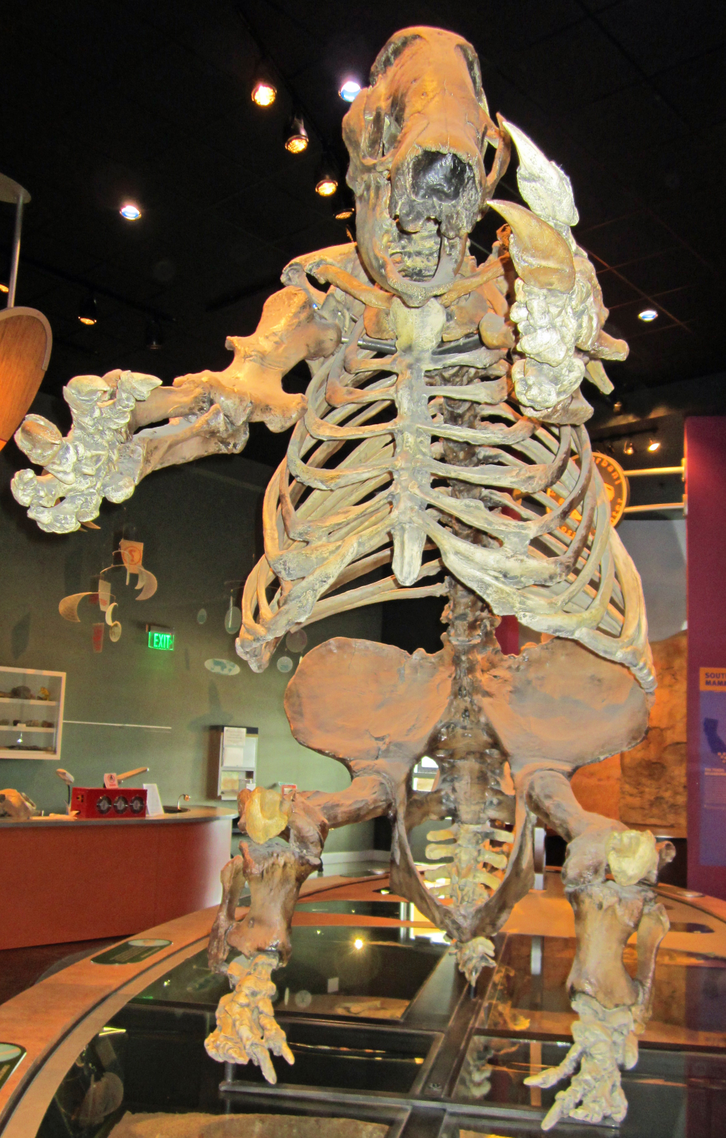
 In many ways, dinosaurs and mammals are very different types of animal. And yet, there are some amazing cases of convergent evolution among them. One of the most bizarre is that of giant ground sloths and therizinosaurs. Like the little slow-moving, tree-dwelling sloths of today, giant ground sloths, like this Paramylodon mounted at Western Science Center, were herbivorous. As a group, sloths are related to armadillos and anteaters. Therizinosaurs, like this Nothronychus mounted at the Natural History Museum of Utah in Salt Lake City, are part of Maniraptora, a group of theropod dinosaurs that also includes predators like Velociraptor and our living dinosaurs, the birds. However, therizinosaurs evolved into big feathery herbivores.
 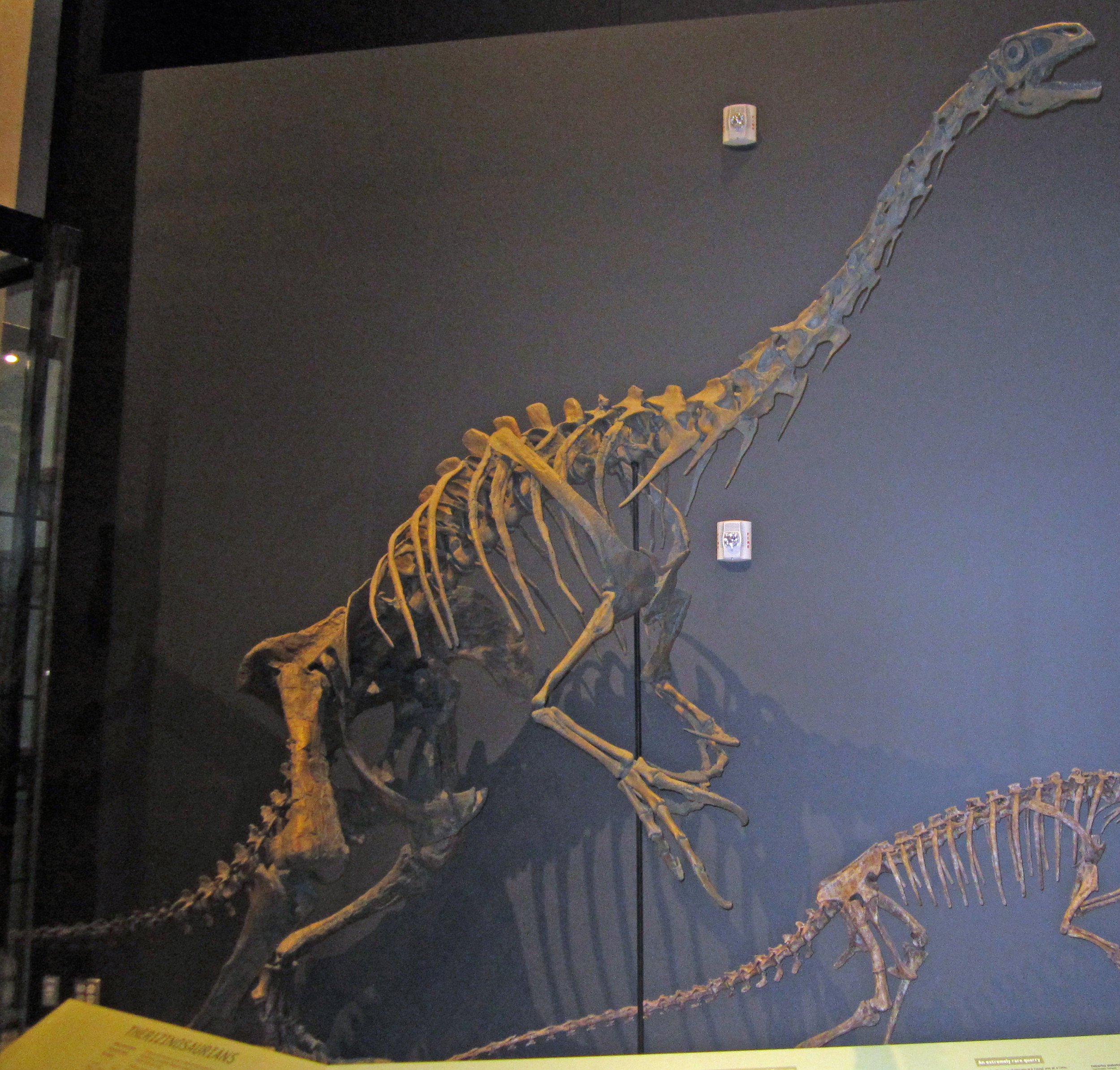 Despite their very different evolutionary origins, giant grounds sloths and therizinosaurs share some remarkable similarities: long arms ending in huge claws, wide bodies to accommodate a long digestive tract for processing plant matter, short robust legs, and short tails. Both groups evolved into large-bodied bipedal plant-eaters. Nothronychus was named in 2001 by my colleagues Jim Kirkland (Utah Geological Survey) and Doug Wolfe (Zuni Dinosaur Institute for Geosciences) - the name means "sloth claw".Another similarity is that both giant ground sloths and therizinosaurs represent migrations into North America from other places. Giant ground sloths evolved in South America and reached North America around nine million years ago. Until the discovery of 90-million-year-old Nothronychus in New Mexico, therizinosaurs were known only from fossils found in Asia. Nothronychus is closely related to the large Asian therizinosaurs and probably represents a migration event into western North America.Post by Curator Dr. Andrew McDonald
Fossil Friday - Paramylodon claw
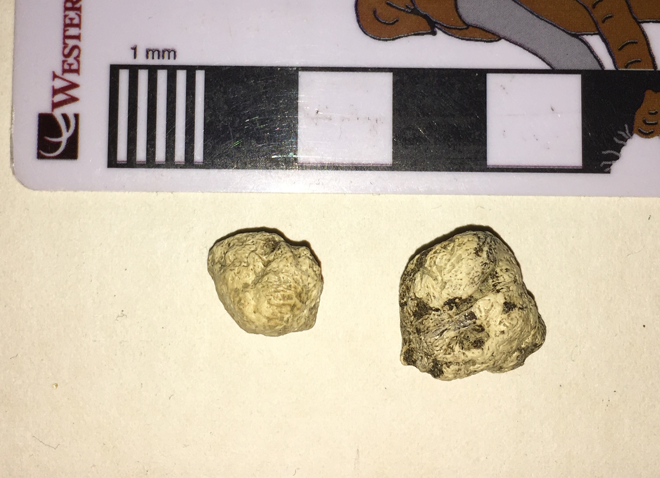
 Sloths are fascinating animals, with all kinds of strange anatomical features. One of their signature characters is their enormous claws. Although bones from the ground sloth Paramylodon are relatively common at Diamond Valley Lake, only a few claws were recovered. Technically, what usually preserves is the last finger bone, technically called the terminal phalanx or ungal. This is shown above from the side, and below from above:
 While we sometimes refer to ungals informally as "claws", it's important to recognize that the ungal is actually the bone that supports the claw. The claw itself is a keratin sheath that fits over the ungal. Keratin is rarely preserved, but there are a few mummified sloths (I don't think Paramylodon is one of them) in which the keratin claw is preserved; it is generally much longer than the ungal.Sloth ungals are always crowd pleasers at outreach events, so we recently molded this specimen and have started producing casts. Now that the molds are completed, the original claw will go back on permanent exhibit in the museum.
Fossil Friday - Paramylodon skull
 Even if a bone is lucky enough to be preserved as a fossil, time is not always kind. There are numerous ways a bone can be altered after burial, including being smushed by the weight of overlying sediment.The lump of bone shown above is actually a nearly complete skull of the ground sloth Paramylodon harlani in dorsal view, collected from the El Casco substation in northern Riverside County. It might be a little difficult to interpret because it has been deformed post-burial. Below is an annotated version:
 The skull is pushed over so that the midline is displaced to the left. This makes features from the right side like the right eye socket easily visible in dorsal view, while hiding the same features from the left side. The entire skull is also flattened, resulting in the wide foramen magnum at the back of the skull.Here's the ventral view:
 And the annotated version:
 Much of the basicranial part of the skull is a big-ole' indecipherable mess, but there are a lot of identifiable features, including all of the tooth sockets.While there are other Paramylodon remains from El Casco, this is the only sloth skull recovered from the site. Even deformed specimens can be significant!
Fossil Friday - sloth mandible
 Greg McDonald's visit last month to look at sloth remains gave us a reason to open our display cases, which include some of our best sloth fossils.One of the exhibit specimens is a nearly complete mandible of Paramylodon harlani, the most common sloth from Diamond Valley Lake. The image is a dorsal view, with anterior to the right. The mandible is nearly complete, if a bit crushed. It's missing the articulation on the right side, part of the anterior end, and all the teeth, but is otherwise in good shape.Paramylodon lower jaws are scoop-shaped and toothless at the tip. This is very different from their distant relative Megalonyx, that has a pair of huge chisel-like teeth at the tip. Even though the teeth are missing, the shape of the sockets also reveals a different cross-section that the more rectangular teeth of Megalonyx. For additional examples of Paramylodon jaws, see our earlier Fossil Friday posts here and here.---This weekend I'll be at the Western Association of Vertebrate Paleontology meeting at Yavapai College in Arizona, presenting on the Mastodons of Unusual Size project and on the Stepping Out of the Past exhibit. Follow @MaxMastodon on Twitter for updates.
Fossil Friday - sloth thoracic vertebra
 Earlier this week, sloth expert Greg McDonald spent several days at the Western Science Center looking at ground sloth material in our collection. I spent that time peering over his shoulder and asking lots of questions. This made the work go more slowly, but it also greatly improved my understanding of ground sloths.At the top is an anterior view of a vertebra from Paramylodon harlani, the most common ground sloth from Diamond Valley Lake. Even though this is a damaged bone, it still shows some important features for understanding sloths.Sloths, along with anteaters and armadillos, are members of the Order Xenarthra, a group that is native to South America. While these animals may seem pretty disparate in their body plans, there are characters that they all share that reveal their relationship to one another. One of the most significant features is reflected in the name of the group; Xenarthra roughly means "alien joint".Most tetrapods have vertebrae that articulate with the vertebrae both ahead of and behind them in the column. Besides the main body of the vertebra (the centrum), there are usually a pair of articulation at the top of the neural canal called the zygapophyses. At the top front edge of the neural arch are the left and right prezygapophyses. The articular surfaces of the prezygapophyses face more-or-less dorsally, and articulate with the postzygapophyses, which are at the top back edge of the neural canal and face ventrally. Below is the dorsal view of a mastodon vertebra featured on Fossil Friday last April, with the prezygapophyses outlined in red (they would normally be symmetrical, but this specimen had been injured):
 Below is an oblique view of the Paramylodon vertebra, looking down from in front (the centrum is partially visible at the bottom of the image):
 Here is the same image, with the prezygapophyses outlined in blue:
 Notice the two articulations outlined in red? Those are the xenarthrous processes (the "alien joints"), which articulate with corresponding points on the posterior edge of the neural canal of the preceding vertebra. The xenarthrous processes are the key feature linking together the various members of the Xenarthra, and are not found in any other group of mammals.Thanks again to Greg McDonald for visiting WSC and giving us so much help with our sloth material.
Fossil Friday - sloth dermal bones
 Next Tuesday evening, Greg McDonald is going to give a lecture at Western Science Center on fossil sloths, so for this week's Fossil Friday we have sloth bones!The tiny bones shown here are actually the most numerous sloth element found at Diamond Valley Lake. These are dermal ossicles, bony nodules embedded in the skin of certain species of ground sloths. Below is another view of the same two specimens:
 Of the three sloth species found at Diamond Valley Lake, only one, Paramylodon harlani, is known to have had dermal ossicles. These two ossicles were found associated with a partial skeleton of Paramylodon from the East Dam (part of the jaw of this individual was featured in an earlier Fossil Friday). In fact, the larger ossicle had rolled into one of the empty tooth sockets in the lower jaw.The presence of dermal ossicles in some ground sloths is a bit surprising. The ossicles are generally assumed to provide armor protection, but they are tiny, and it's not clear how extensive they were. While several patches of preserved Paramylodon skin have been found with numerous embedded ossicles, we don't seem to find enough ossicles to cover the entire, or even most, of the body. It's also curious that most ground sloths seem to have gotten along perfectly well without dermal armor, including Megalonyx, which was close to the same size as Paramylodon, overlapped with it in time and space, and was even more widely ranging. Dermal armor is common in some other groups within the Xenarthra (the order that includes sloths), such as the armadillos and glyptodonts. So it's possible that the armor in Paramylodon is a relict, a holdover from some earlier sloth ancestor. But this would mean the earliest sloths should have had armor (as far as I know this is not the case), and that armor was subsequently lost in almost every sloth lineage except for a few species. So for now the origin and function of Paramylodon dermal ossicles remain a bit mysterious.
Fossil Friday - sloth jaw
 Yesterday was International Sloth Day (with sloth in this case being a noun, not an adjective)! That's a nice day to celebrate at Western Science Center, because Diamond Valley Lake is the only locality in California with three different species of ground sloths. By far the most common sloth at DVL is Harlan's ground sloth, Paramylodon harlani. Shown above is a Paramylodon partial lower jaw. This is the anterior end of the right dentary seen in dorsal view, with anterior to the left. The long straight edge at the lower left is the mandibular symphysis, where the right and left dentaries attach to each other (essentially the chin). The bases of two teeth are visible at the upper right.Here's a lateral view:
 This time the anterior end is on the right. In this view it's clear that the teeth are broken off, and barely protrude above the bone. This is presumably breakage that occurred after the animal died, since the breaks are jagged.This fragment makes an interesting comparison with one of the other DVL sloths, Megalonyx jeffersonii. Paramylodon seems to have a longer, more slender jaw than Megalonyx and lacks the latter's enlarged, chisel-like incisors. That's not entirely surprising; while Paramylodon and Megalonyx are both sloths, they are only distantly related to each other.
Fossil Friday - sloth ulna
 Last Saturday the Western Science Center was visited by Eric Scott from The Cooper Center and high school student Santiago Hernandez. They came with a specific goal in mind, to look at fossil horses from a small, understudied collection, called the Harveston Collection, from western Riverside County.While the Harveston Collection is dominated by horses, there are several other kinds of animal present, including bison, camels, deer, and mammoths. As we examined each drawer looking for horses, Eric noticed the bone shown above, which was labeled in the field as a juvenile bison humerus. We both immediately agreed that this bone was not a humerus, as is lacked the distinctive articulations found in the humerus at both the proximal and distal ends. After a few minutes of debate, we decided that the bone looked more like a damaged ulna (a bone from the forearm), perhaps from a ground sloth. Conveniently, the ground sloth Paramylodon was common at Diamond Valley Lake, and we have a cast mounted skeleton on exhibit. So, with the horses temporarily forgotten, we rushed off to the exhibit hall to compare the Harveston specimen to Paramylodon (thanks to Santiago for the photo):
 It turned out that our suspicion was correct; allowing for breakage, the bone is a perfect match for the right ulna of Paramylodon. This was a neat discovery, as this is the only sloth bone currently identified from the Harveston Collection (although I think sloths had previously been found at nearby localities). There are new discoveries to be found in museum collections all over the world.
Fossil Friday - sloth vertebra
 One of the most enjoyable things about writing this blog is that I have the chance to learn about the anatomy of animals that are relatively unfamiliar to me. While I've done a little work on sloths in the past, their somewhat unusual skeletal anatomy can be tricky for someone who has mostly worked on other animals.Sloths and their relatives have unique articulation features (the xenarthrous processes) in their posterior thoracic and lumbar vertebrae. In order to understand these better, I asked Darla, the WSC Collections Manager, to search our collections database for lumbar vertebrae from our most common sloth, Paramylodon harlani. It turns out that, according to the database, we only have one reasonably well preserved Paramylodon lumbar that isn't already on exhibit, and she pulled that specimen out for me to examine. Shown above is the anterior view, and below is posterior:
 The vertebra isn't complete. The entire neural spine is missing, as are the posterior articular surfaces (the postzygopophyses), and there is damage to the centrum. The transverse processes are also broken off, as is more apparent in right lateral view:
 In dorsal view (below) two big anterior troughs are visible. These would have articulated with the postzygopophyses of the vertebra in front of this one.
 I struggled for some time trying to understand this vertebra. I was completely unable to locate the xenarthrous processes, and the transverse processes made no sense when I compared this specimen to a cast Paramylodon lumbar vertebra. After spending some time with references, especially Stock (1925), I figured out why I was having such difficulties; the vertebra is not a lumbar, but is in fact a caudal (tail) vertebra that was misidentified on the label! This is pretty easy to spot when compared to dorsal view figures of Paramylodon caudals from Stock:
 To my chagrin, once I finally realized this was a caudal vertebra, I saw that there were plenty of indicators I had overlooked (like articulations for the haemal arches) that should have told me right away that this was a caudal. But starting with the assumption that it was a lumbar made it slower for me to to recognize contrary data for what it was.I should also note that, while misidentifications do occasionally slip into databases, or even publications (I've done that too!), it's not as common as you might think. The Diamond Valley Lake collections are, in my opinion, extremely well identified. I've now looked in some detail at more than a thousand different bones and teeth in the DVL collection. This vertebra is only the third instance in which I've disagreed with the initial identification. There have also been at least four other instances where I thought there was a misidentification, but closer examination convinced me that the original ID was correct. This constant reevaluation and self-correction is at the core of scientific methodology, and is what makes it such a powerful tool for understanding the world.Reference:Stock, C. 1925. Cenozoic Gravigrade Edentates of Western North America with Special Reference to the Pleistocene Megalonychinae and Mylodontidae of Rancho La Brea. Carnegie Institute of Washington Publication 331, 206 p., 47 pls.
Fossil Friday - Harlan's ground sloth femur
 There are three different species of ground sloths known from the Pleistocene Diamond Valley Lake fauna. By far the most common of the three is Harlan's ground sloth, Paramylodon harlani. For this week's Fossil Friday we have a Paramylodon femur.This is the right femur, seen from the back (posterior view), with the distal end on the right. The articular surfaces for the knee joint are visible on the right, but the proximal end of the bone is not preserved so the ball joint where the femur attaches to the pelvis is missing (it would have been at the lower left corner of this image). Even though the bone is crushed, it has maintained its shape pretty well. Like many of the larger bones in the WSC collection, this one has only been prepared on one side.I've always found sloth femora fascinating, because they have a really strange shape when compared to most of the other mammals we find in Pleistocene deposits in North America. They appear to be very wide for their length, and generally kind of overbuilt for their size. To get an idea, compare this femur to the juvenile mastodon femur I wrote about a few months ago. These animals were probably roughly the same size, but the difference in their femoral proportions is striking.———On another note, we are busily preparing for tomorrow's Inland Empire Science Festival. If you're in Southern California we hope you can make it out to the Western Science Center tomorrow for this exciting event.
Fossil Friday - Harlan's ground sloth jaw
 For this week's Fossil Friday we have a partial lower jaw of Harlan's ground sloth, Paramylodon harlani, collected near the eastern end of Diamond Valley not far from the museum's current location.This particular fragment is the back half of the left dentary (the left and right dentaries are the bones that make up the lower jaw in mammals). The image above is the lateral (side) view with the front to the left. The small projection sticking out on the middle of the back edge is the mandibular condyle, the part of the jaw that forms the joint with the rest of the skull.Here's the medial view of the same jaw:
 And here's the dorsal (top) view:
 In this view we can see that, while the teeth are missing, the tooth sockets for the 3rd and 4th molars are preserved, as well as the back and inner edges of the socket for the 2nd molar. The mandibular condyle also appears much larger from this angle, because while it is relatively short vertically it is quite wide transversely (from side-to-side).There are several different species of sloths known from the Diamond Valley Lake region and that are represented in the WSC collections. Paramylodon harlani is by far the most common, making up over 93% of the individual sloth bones from the valley (Springer et al. 2010).Reference:Springer, K., E. Scott, J. C. Sagebiel, and L. K. Murray, 2010. Late Pleistocene large mammal faunal dynamics from inland southern California: the Diamond Valley Lake local fauna. Quaternary International 217:256-265.
