

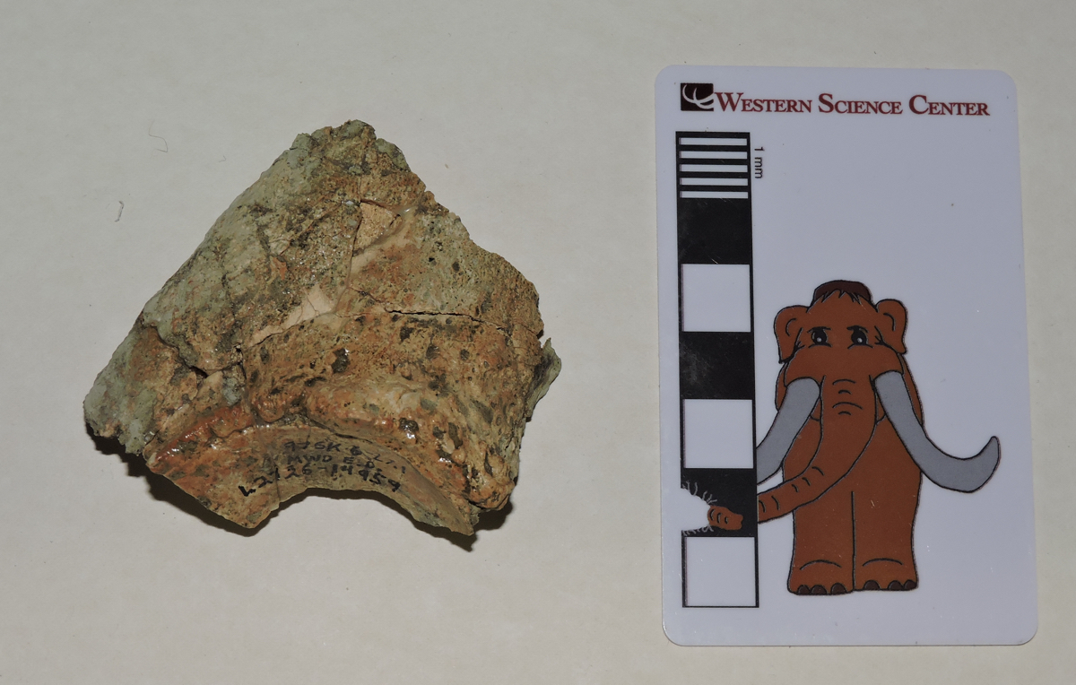
 Taphonomy is the subset of paleontology that examines how an organism gets preserved as a fossil. Taphonomists consider all kinds of different features, including the structure and composition of the organism, how and where it died, rates of sedimentation, the quantity and chemical composition of groundwater in the area, and countless other variables. Understanding taphonomy can tell us a great deal about an organism and its environment, and it's something to be considered when looking at unusual preservation.The specimen shown here is the partial palate of a mastodon, seen in palatal view (looking up at the roof of the mouth). All that remains are the crowns of the teeth and few bits of the maxillary and palatine bones. I'm not sure why this specimen preserved this way, but there are some possibilities to consider. By far the best preserved portions are the crowns of the teeth. Tooth enamel is generally the hardest and most chemically resistant part of the vertebrate body, so it's not entirely surprising to see teeth surviving while the rest of the skeleton deteriorates.This specimen has both left and right teeth preserved, in their correct life positions relative to each other, even though most of the bone between them is gone. That means the upper jaw survived as a unit for at least some period of time. Perhaps the skull was sitting on the ground in such a way that the upper jaw was buried but the rest of the skull was exposed and eroded away. Maybe when the animal was rotting or scavenged, the maxillae separated from the rest of the skull. Or maybe the skull was largely intact when it was buried but groundwater mostly dissolved the bones and left the teeth behind (we have a horse in the collection that may have experienced this).As for the mastodon itself, the specimen is oriented with anterior to the left, so the left teeth are at the top and the right teeth are at the bottom. Portions of the crowns of both upper 2nd molars are preserved, and the left one is almost complete. The crowns of both upper 3rd molars are also preserved. The 2nd molars show heavy wear, but the 3rd molars show very little wear, and only on the anterior part of the tooth. This suggests a mastodon that was probably in its early 20s when it died.---As a reminder, tomorrow is the 2nd Annual Inland Empire Science Festival at the Western Science Center. Come to the museum to for tons of science-themed activities, lectures, and exhibits. Student, youth, and under-4 admissions will receive a coupon for a free replica Tyrannosaurus tooth, molded from a specimen on exhibit at the museum.
Fossil Friday - mole claw
 Most of the fossils found at Diamond Valley Lake came from relatively small animals. While rodents dominate the small mammal fauna, animals other than rodents are also present, including moles.Above is a partial claw from a broad-footed mole, Scapanus latimanus. Moles are in the Order Eulipotyphla, a group that includes a variety of animals that used to be placed in the Insectovora (it turns out quite a few of the insectivorans were not closely related to each other, and the grouping is rarely used anymore).Moles are burrowing mammals, digging tunnels through the soil with large, powerful front feet, as can be seen in these examples of related eastern moles (Scalopus aquaticus) from the North Carolina Museum of Natural Sciences and the Museum of Osteology, respectively:
 Moles can live in a variety of conditions, but they are restricted to soils with enough moisture and the correct texture for making their burrows (they don't live in soil that's too sandy, for instance), and to areas where there is enough food to sustain them. Like many small mammals moles are voracious predators that eat a significant percentage of their body weight each day, so they require a steady supply of earthworms, insect larvae, snails, and other burrowing invertebrates. Many of these invertebrates have a low preservation potential, but the presence of moles in a deposit hints at their presence.
Fossil Friday - ram's horn snail
 During the Pleistocene, Diamond Valley seems to have been quite a bit wetter than it is now (at least, than it was before it was turned into a reservoir). A fair number of organisms associated with standing water were collected in these deposits, including snails.The three shells shown above are examples of a species of ram's horn snail, the ash gyro Gyraulus parvus. Like most of the snails from the Diamond Valley Lake deposits, they are tiny; the black square they're sitting on is 1 cm on each side.Modern G. parvus is a widespread species in North America, and is found in a variety of freshwater habitats that involve standing or slow-moving water. According to at least some sources they seem to prefer stable (rather than intermittent) bodies of water. Ash gyros live on the bottom of the pond or attached to water plants, where they feed on benthic diatoms (single-celled algae).
Fossil Friday - sloth vertebra
 One of the most enjoyable things about writing this blog is that I have the chance to learn about the anatomy of animals that are relatively unfamiliar to me. While I've done a little work on sloths in the past, their somewhat unusual skeletal anatomy can be tricky for someone who has mostly worked on other animals.Sloths and their relatives have unique articulation features (the xenarthrous processes) in their posterior thoracic and lumbar vertebrae. In order to understand these better, I asked Darla, the WSC Collections Manager, to search our collections database for lumbar vertebrae from our most common sloth, Paramylodon harlani. It turns out that, according to the database, we only have one reasonably well preserved Paramylodon lumbar that isn't already on exhibit, and she pulled that specimen out for me to examine. Shown above is the anterior view, and below is posterior:
 The vertebra isn't complete. The entire neural spine is missing, as are the posterior articular surfaces (the postzygopophyses), and there is damage to the centrum. The transverse processes are also broken off, as is more apparent in right lateral view:
 In dorsal view (below) two big anterior troughs are visible. These would have articulated with the postzygopophyses of the vertebra in front of this one.
 I struggled for some time trying to understand this vertebra. I was completely unable to locate the xenarthrous processes, and the transverse processes made no sense when I compared this specimen to a cast Paramylodon lumbar vertebra. After spending some time with references, especially Stock (1925), I figured out why I was having such difficulties; the vertebra is not a lumbar, but is in fact a caudal (tail) vertebra that was misidentified on the label! This is pretty easy to spot when compared to dorsal view figures of Paramylodon caudals from Stock:
 To my chagrin, once I finally realized this was a caudal vertebra, I saw that there were plenty of indicators I had overlooked (like articulations for the haemal arches) that should have told me right away that this was a caudal. But starting with the assumption that it was a lumbar made it slower for me to to recognize contrary data for what it was.I should also note that, while misidentifications do occasionally slip into databases, or even publications (I've done that too!), it's not as common as you might think. The Diamond Valley Lake collections are, in my opinion, extremely well identified. I've now looked in some detail at more than a thousand different bones and teeth in the DVL collection. This vertebra is only the third instance in which I've disagreed with the initial identification. There have also been at least four other instances where I thought there was a misidentification, but closer examination convinced me that the original ID was correct. This constant reevaluation and self-correction is at the core of scientific methodology, and is what makes it such a powerful tool for understanding the world.Reference:Stock, C. 1925. Cenozoic Gravigrade Edentates of Western North America with Special Reference to the Pleistocene Megalonychinae and Mylodontidae of Rancho La Brea. Carnegie Institute of Washington Publication 331, 206 p., 47 pls.
Fossil Friday - Bison latifrons horn core
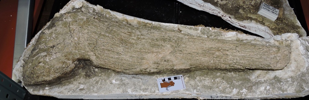 As I've mentioned in previous posts, there were two different species of Bison present at Diamond Valley Lake, the larger, long-horned Bison latifrons and the (relatively) smaller, shorter-horned Bison antiquus. These species are closely related, and it's often difficult to distinguish between them when dealing with fragmentary remains. But sometimes there's no doubt.Above is the right horn core from Bison latifrons. It has only been partially prepared, and is still in its field jacket. It's shown in dorsal view, with medial to the left and lateral to the right.Like other bovids, bison horns have a bony core that grows from the frontal, one of the bones that makes up the roof of the skull. The lump of bone on the left is part of the frontal. As large as the core is, it's incomplete; at least several centimeters are broken off from the tip. In life, the bony core would have been covered by a keratin sheath, making the horn even larger. And, of course, there was another horn on the left side. All told, it makes for an impressive horn spread, as shown in this cast from the Sam Noble Oklahoma Museum:
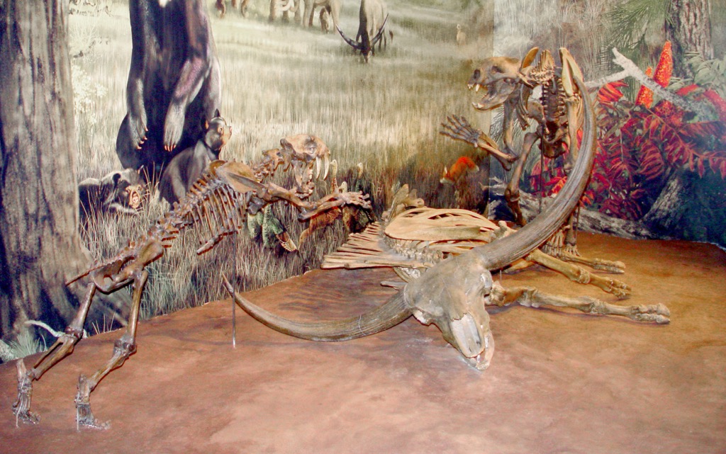
Fossil Friday - Homotherium jaw
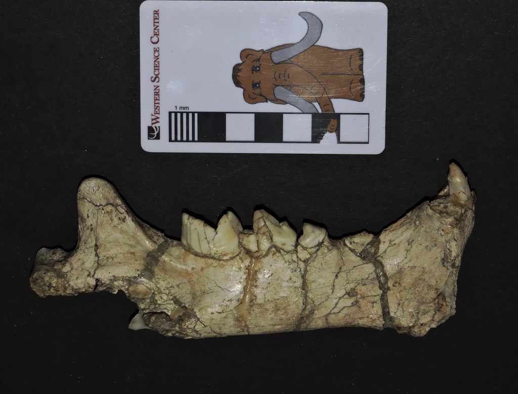 A basic tenet of ecology is that as you move higher up a food chain, the biomass (and usually number of individual animals) will get smaller. Each trophic level in a food chain gets its energy from the level below, and since there is never a perfect 100% energy transfer there is always less energy available at higher levels. This has a profound impact on the fossil record; in general, "top-of-the-food-chain" carnivores are exceptionally rare as fossils, because they were also rare as living animals. (If you spend time outdoors, think about how many squirrels, rabbits, and deer you've ever seen, compared to the number of coyotes and bobcats.) While there are occasional exceptions such as the predator trap at Rancho La Brea that bias the fossil record in favor of carnivores, at most localities large carnivores are rare.The fossils Southern California Edison El Casco Substation site are dominated by rodents, horses, and other herbivores, but a handful of large carnivores were also discovered. One of the most fascinating is the "other" Ice Age sabertooth cat, Homotherium.A number of bones from one individual of Homotherium were collected at El Casco, including the right dentary (lower jaw) shown here. At the top is the lateral view, and below is the medial view:
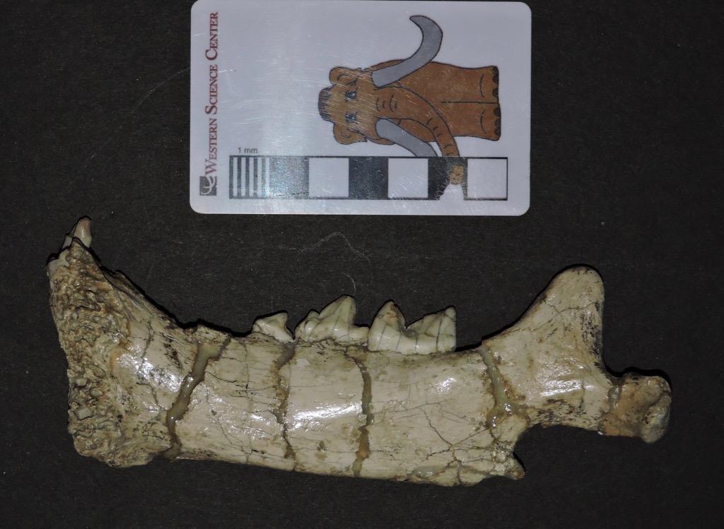 The dentary is largely complete, although there is some damage at each end, and several teeth are broken. There is a flange on the bottom edge at the front of the jaw; this is a common feature in sabertooth cats (and some other saber-toothed animals) to help protect the long upper canine teeth.Below is a dorsal view, showing the occlusal side of the teeth:
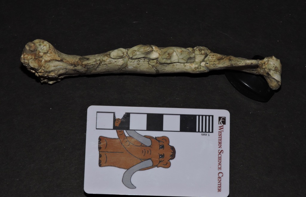 A labeled version of the same image:
 Cats, including Homotherium, have a highly specialized dentition. Each half of the lower jaw includes 3 incisors and an enlarged canine. Most of the premolars and molars have been lost in advanced cats, with only the 3rd and 4th premolars and the 1st molar remaining. These are modified into sharp blades that, when occluding with the upper teeth, act like a pair of scissors for slicing meat. Cats lack a grinding molar in the back of the mouth like the kind seen in dogs, and as a result are much less likely to gnaw on or attempt to crack open the bones of their prey.One of the unusual features of Homotherium is its enlarged incisors. It's not obvious in this specimen because the canine tooth is broken, but the lower incisors were almost as large as the canine, and protrude more from the front of the mouth than in most other cats. There's a beautiful illustration of Homotherium by Mauricio Anton that shows details of the unusual dentition.Homotherium was a large, tiger-sized cat with rather short back legs compared to the front legs, as can be seen in the reconstructed skeleton at the Texas Memorial Museum show below. Several different species of Homotherium are known from both the New World and the Old World, and they probably filled a variety of different ecological niches. There is at least one site in Texas that suggests that a Homotherium population there may have had a preference for juvenile mammoths as prey items, but mammoths are exceptionally rare in the sediments that produced the El Casco Homotherium.
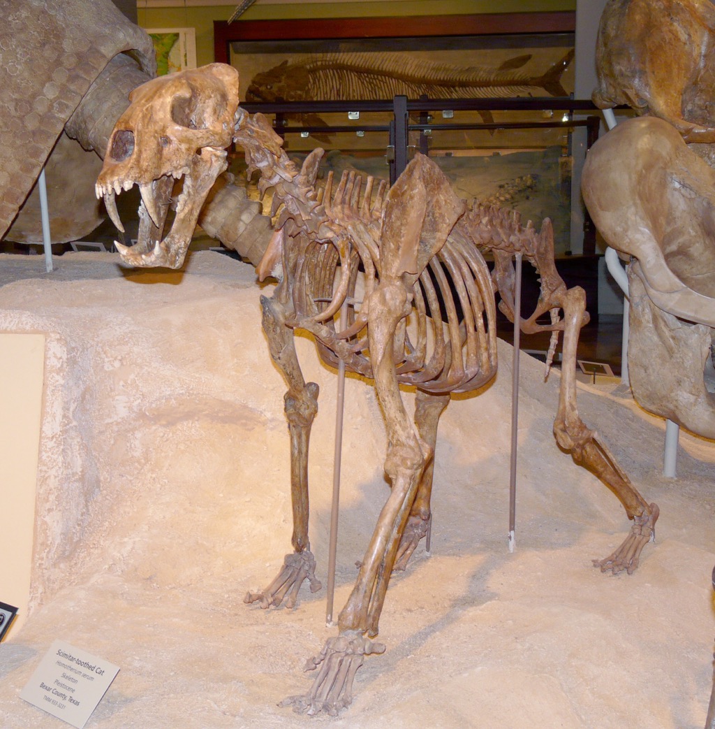
Fossil Friday - mastodon pelvis
 When the permanent exhibits were being installed at WSC, several high-profile specimens were molded and cast for the displays, including the large mastodon nicknamed Max. It turned out that Max's pelvis was too fragile to withstand molding, so while the pelvis is on exhibit it was the only recovered element of Max's skeleton that wasn't reproduced.The mammalian pelvic girdle is a complex structure made up of multiple bones that are generally separate in young animals but mostly fuse with age. All of the various pelvic bones are preserved in Max, although some of them are incomplete.The image above shows the pelvis in close to dorsal view, maybe somewhat posterodorsal (above and behind), with a Max scale bar for scale. It is rotated so that anterior is toward the top of the image. Below is a marked-up version of the same photo:
 The largest bone, outlined in blue, is the ilium (only the bones on the right side and the midline have been color-coded). The ischium is outlined in red, and the pubis, partially hidden under the overlying bones, is yellow. The bulge on the side where the ilium and ischium meet is the hip socket, or acetabulum. The actual socket opens to the side and down, so it's hidden in this view. The pubis also comes up to the bottom of the acetabulum, but that is also hidden in dorsal view.The left and right ilia are separated by, and fused to, a structure called the sacrum, which is made up of a series of fused sacral vertebrae, outlined in purple above. Max appears to have four sacral vertebrae; the number can vary on an individual basis, and some mastodons have five. The first two tail vertebrae (caudal vertebrae) are also present, outlined in dark blue. It's not clear if they are actually fused to the sacrum, or simply interlocked with it. Excluding the tail vertebrae, Max's pelvis is therefore made up of 10 bones: the left and right ilia, left and right ischia, left and right pubes, and four sacral vertebrae.Besides serving as the attachment points connecting the legs to the body, the pelvic girdle supports the back of the abdominal cavity. The digestive tract exits the abdominal cavity through the urethra and anus, so the pelvic bones are arranged in a tube to allow for this, as well providing a path for the birth canal in females. The sacrum forms the top of this tube, while the ilia and ischia make up the sides and pubes make up the bottom. This is more obvious is we look at the pelvis from below and behind (posteroventrally) - exactly where you don't want to stand with an elephant that has recently eaten or that's about to give birth:
 Below are two more views of Max's pelvis, to give a better idea of the arrangement of the various bones. First, an oblique view from above the right side (anterior is to the lower right):
 And above the left side (anterior is to the lower left):
 Notice in each of these views, and especially in the marked-up image, that the boundaries between the various bones are not immediately obvious. Max, while not elderly, was a fully mature mastodon (probably over 30 years old), so these bones were solidly fused to each other. In a younger mastodon the boundaries between the bones would be more obvious.Max's pelvis only has a partial support cradle, so unfortunately we can't flip it over to see the ventral side. Even so, we can see quite a lot in the views that are available.
Fossil Friday - fence lizard jaw
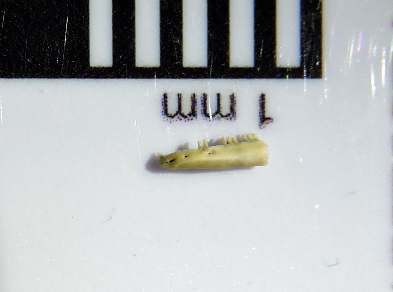 Lizards are a common site today across southern California, and that was true in the Pleistocene as well. Large numbers of lizard bones were recovered from Diamond Valley Lake sediments, including the one shown here.This is a partial left lower jaw from a lizard of the genus Sceloporus, most likely a western fence lizard (Sceloporus occidentals). These small lizards live all over California today, and are often sunning in my driveway when I get home from work:
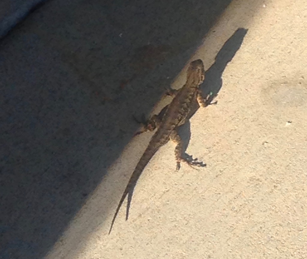 As I mentioned in an earlier post, mammals have lower jaws that consist of a single bone, the dentary, on each side. In other vertebrates the dentary is one of several bones that make up the lower jaw. This particular specimen includes the anterior part of the left dentary, while the back part of the dentary and the other lower jaw bones are all missing. The view at the top is a lateral (side) view, with anterior to the left. Below is a medial view, anterior to the right:
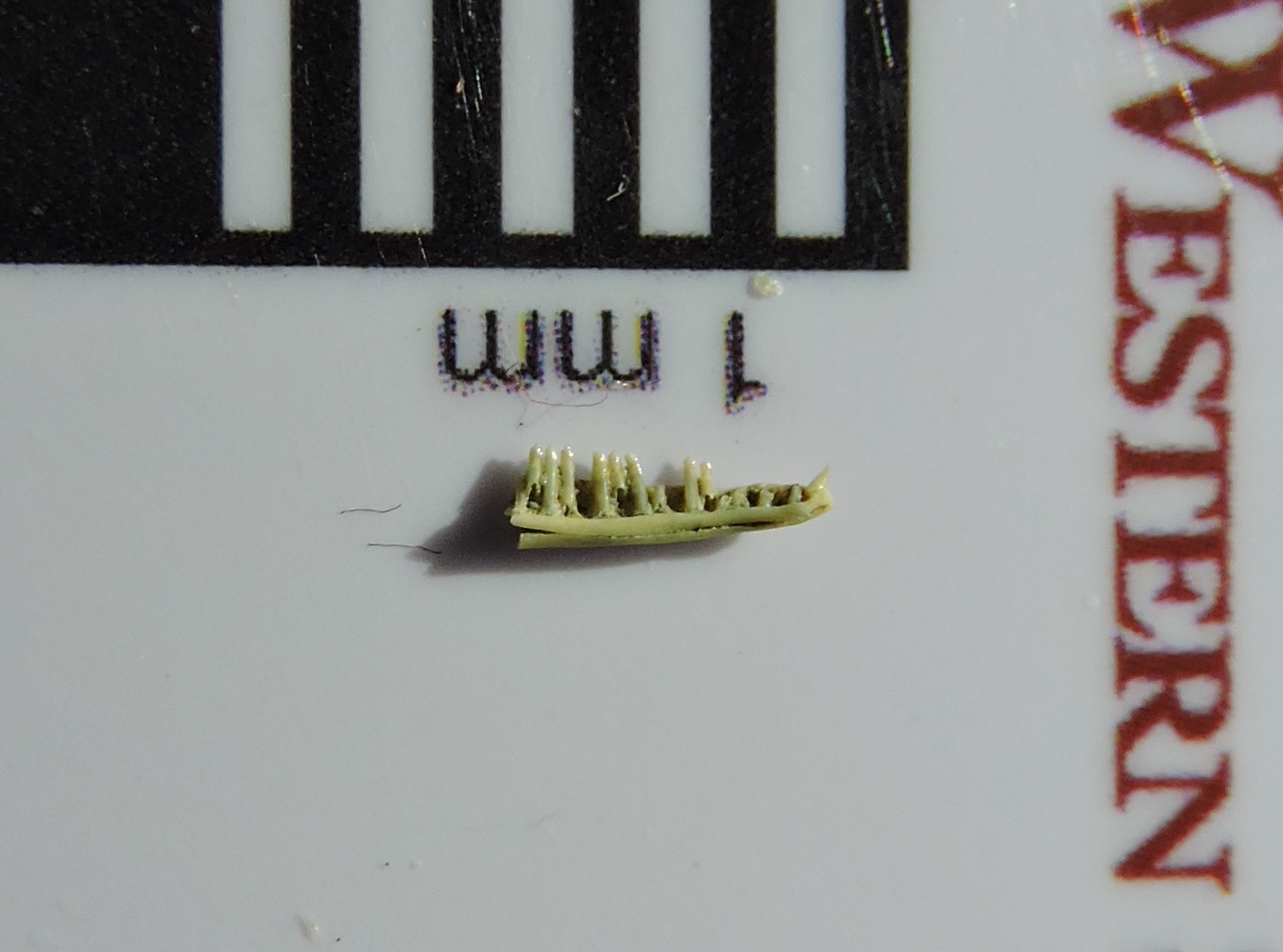 In this view we can more clearly see there are nine complete teeth preserved, and the bases of at least five others.Here's a dorsal or occlusal view, anterior to the left:
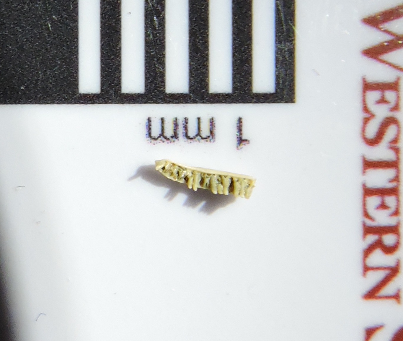 Fence lizards are widespread today and live in a variety of environments, so the presence of fence lizards doesn't really give us any information about the paleoenvironmental conditions at Diamond Valley Lake during the Pleistocene. But if you traveled back to the Pleistocene and could pull your eyes away from the mammoths, mastodons, and other megafauna long enough to look at the small animals among the shrubs and rocks, what you would see would look a lot like modern-day California.
Fossil Friday - bison ungal
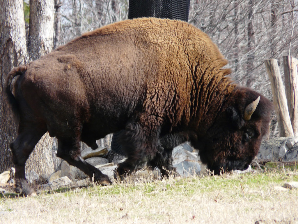
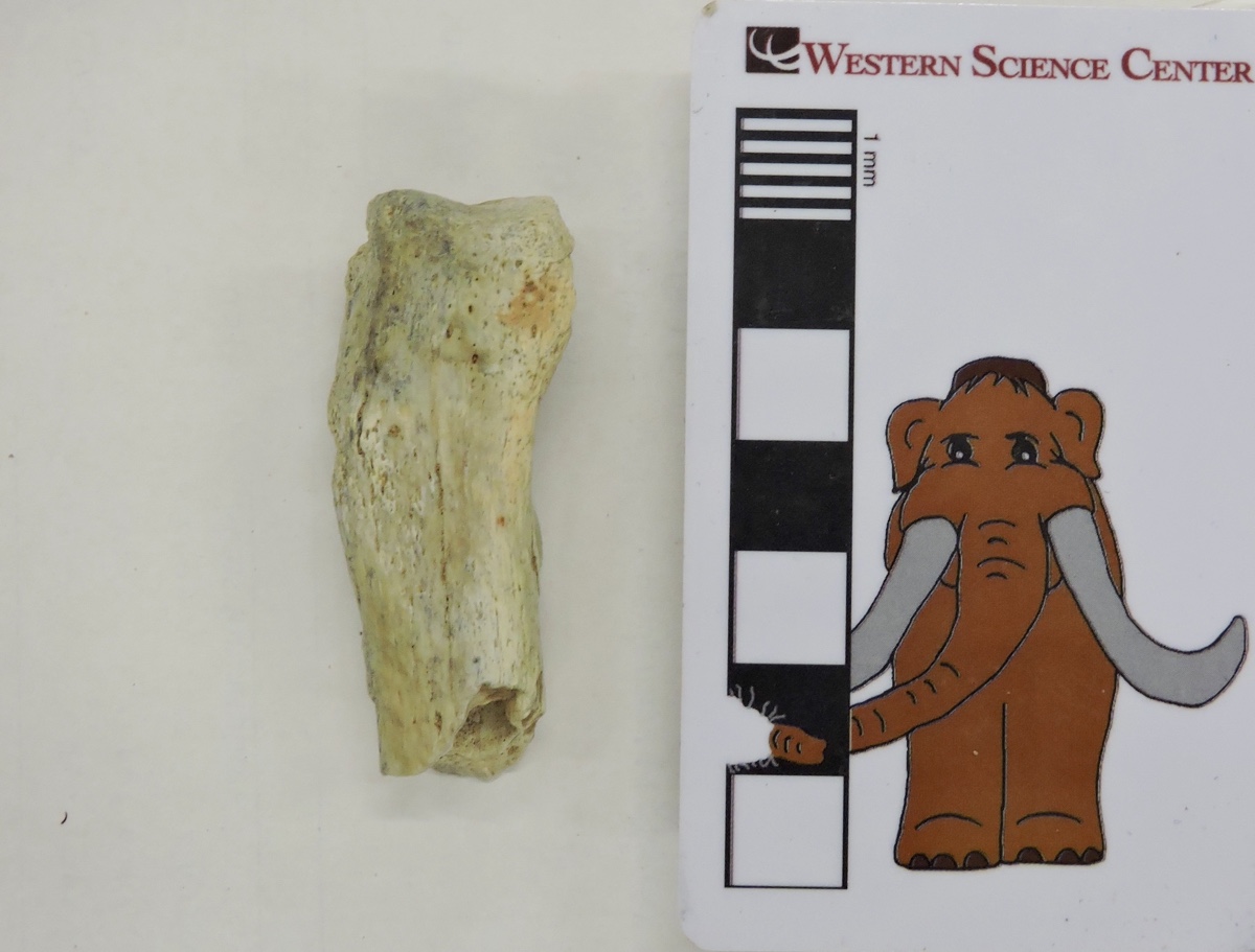
 Hooves are present in several groups of mammals, including the Artiodactyla, the diverse order that includes cattle (bovids) and an enormous range of other animals. Bovids, including bison, have only two toes on each foot, with a hoof at the tip of each toe, as is clear in the photo below of a bison considerately presenting his hooves for inspection:
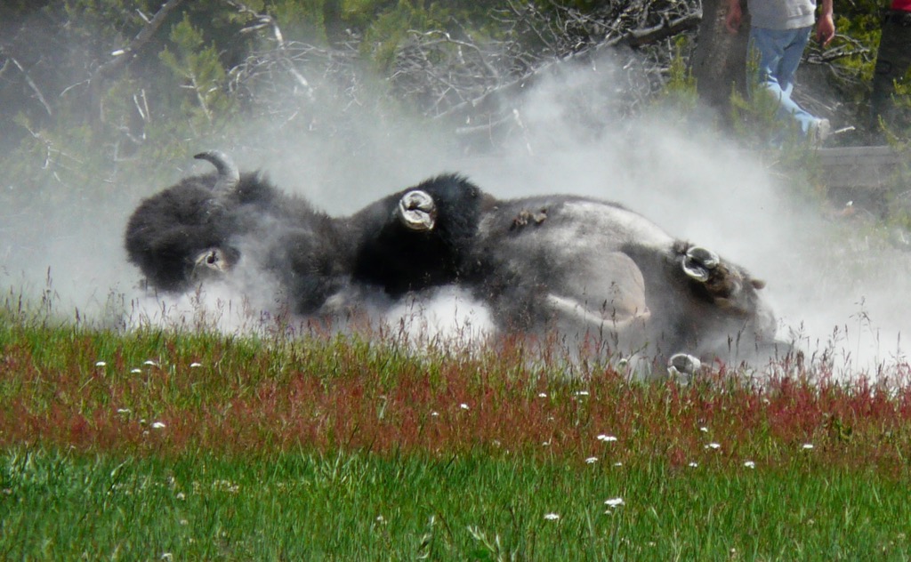 The outside of the hoof is made largely of keratin proteins, but inside is a bone. This is the last finger or toe bone, known as the terminal phalanx or ungal. The ungal shown above appears to be from left manual digit IV, or the outside toe on the left forefoot. This specimen was collected from the East Dam of Diamond Valley Lake, not far from where the Western Science Center is now located.
Fossil Friday - bison lower jaw
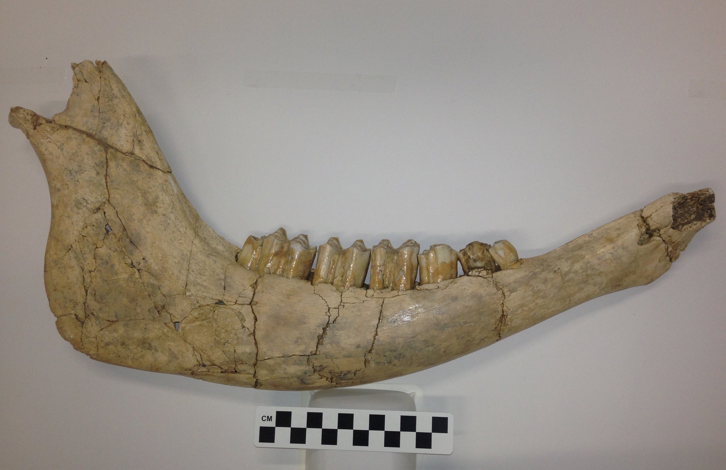 While the Diamond Valley Lake deposits didn't produce anything close to a complete skeleton of a bison, their bones are still among the most common from large animals in the valley, and many of them are well preserved.This is the right side of the lower jaw of Bison antiquus, the smaller of the two Pleistocene bison species from California. Mammals are unique in that each half of the lower jaw is made up of a single bone, called on he dentary, so above is the lateral view of the right dentary. Here's the medial view of the same bone:
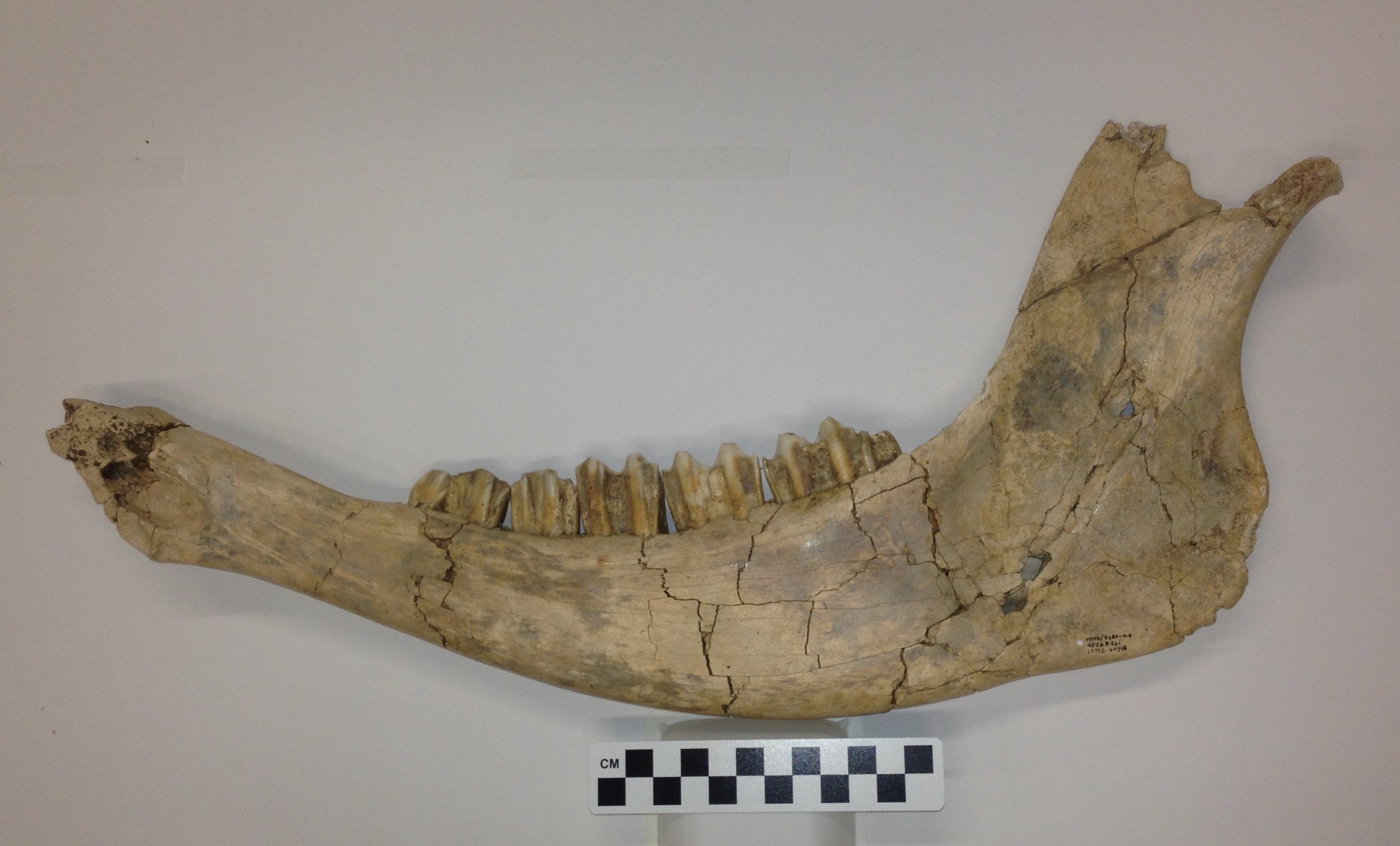 The bone is missing the anterior tip and part of the coronoid process (at the very top), but otherwise is essentially complete. Since the anterior end is absent, the incisor teeth located there are also missing, but otherwise we have the complete dentition. This includes the 2nd, 3rd, and 4th premolars (bison have lost the 1st premolar), and the 1st, 2nd, and 3rd molars. Below is an occlusal view, showing the chewing surfaces of the teeth:
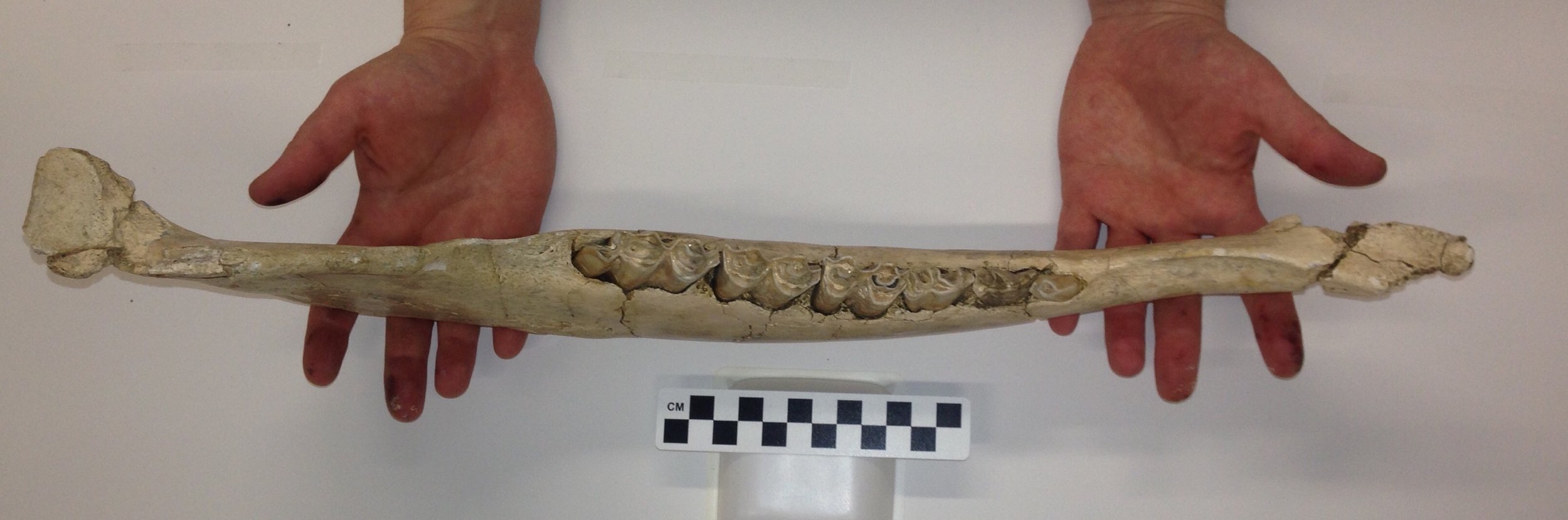 This is a complete set of permanent teeth, so this jaw represents an adult animal, although the moderate wear suggests that it was more likely middle-aged than elderly.This dentary is currently on display at the Western Science Center in the Stories from Bones exhibit.
Fossil Friday - mastodon rib
 Here at Valley of the Mastodon, we're ringing in the new year with a rib from our namesake animal. Ribs don't often get a lot of attention, but there sometimes is information that can be gleaned from them.The left rib shown above is incomplete, with portions of both ends of the bone missing. The proximal end, closest to the vertebrae, is to the right. This rib is large enough to be from an adult or near-adult animal, but it's impossible to estimate the age with any certainty. Ribs do have an epiphysis at the proximal end that is unfused in young animals, but that area's not preserved in this specimen.So what's going on with the proximal end of this rib? Here are some closeups from various angles:

 A swollen region around the bone, pits and bone spurs on the surface...these are all indications of an injury or other pathology. As with many of the specimens in our current "Stories from Bones" exhibit this bone shows a healing response to some type of trauma. It's difficult to say what the trauma was. The bone could have been broken in an attack by a predator, and while there are no indication of bite marks to corroborate this it's still a possibility. The rib could have been broken in a fall or some other accident, or in a fight with another mastodon. Infections and certain diseases can also sometimes cause this type of response in a bone, but we are definitely looking at something that happened to this mastodon while it was still alive.
Fossil Friday - deer antler
 With Fossil Friday falling on Christmas, I would have liked to post a picture of a fossil reindeer (Rangifer tarandus). But most of the vertebrate fossils at Western Science Center are from Riverside County, and while reindeer (or caribou, as they're called in North America) did make it as far south as Georgia during the Ice Age they apparently never made it to southern California. So instead I've gone with the closest relative to reindeer in our collection.Rangifer is a member of the family Cervidae, the deer. A unique and highly visible feature of cervids is the presence of antlers in males in almost all extant species (a few species don't grow antlers, and in reindeer the females also grow them).Shown above is the base of an antler from the Early Pleistocene deposits in San Timoteo Canyon, found during the Southern California Edison El Casco Substation project. This is the left antler seen in lateral view, with anterior to the left. The bone at the base is a fragment of the frontal, which makes up part of the braincase. The cylindrical shaft angling back from the frontal is called the pedicle, while the rugose portion at the tip is the coronet. Here's a medial view of the same fragment:
 Antlers are unique in that, even though they are made of bone, they are shed and regrown periodically, usually every year. The pedicle is the only permanent part of the antler. In contrast, other mammals with comparable head gear, such as bovids (cattle and antelopes), have horns that consist of a bony core covered with a keratin sheath and that are never shed (pronghorns shed the keratin sheath but not the bony core).(As an aside, antler growth is initiated by hormones stimulating specialized connective tissue that covers the pedicle. In researching this blog post, I was surprised to find that injuries to the skin in other parts of the head can result in antler growth at the sites of the injuries, so that occasionally a deer will grow extra antlers (Bubenik and Hundertmark 2002)!)When I first looked at this specimen, I was struck by its size; it's much larger than the white-tailed deer (Odocoileus virginianus) I frequently saw back east. I briefly wondered if it might be from an elk (Cervus elaphus), another species that, like reindeer, apparently never made it to southern California. We happen to have a modern elk in the WSC collection, and a quick comparison showed that, large as it is, our fossil specimen is much smaller than a mature elk:
 This specimen is probably close to the upper end of the size range for a mule deer, Odocoileus hemionus, which does live in southern California. There may also be extinct species of Odocoileus from the Early Pleistocene that are possibilities, but with only an antler fragment it may not be possible to make a definite identification of this specimen beyond the family or genus. Reference: Bubenik, G. A. and Hundertmark, K. J., 2002. Accessory antlers in male Cervidae. Zeitschrift für Jagdwissenschaft 48:10-21.
Fossil Friday - mastodon molar
 I've been looking at a lot of mastodons lately, so they're likely to show up more and more on Fossil Friday. Today's entry is an upper left 1st molar, collected from the West Dam at Diamond Valley Lake.The tooth is shown above in labial view (the side of the tooth on the outside of the mouth, literally closest to the lips), with anterior to the right. Of course, since it's an upper tooth, I really should have photographed it with the crown at the bottom.Here's the lingual side (literally, closest to the tongue):
 Notice that the pointed parts of the crown (called lophs) are more worn in this view. In mastodons (and in many other mammals with grinding molars) the crowns of upper teeth will wear more rapidly on the more the labial side. The reverse is generally true of the lower teeth.Here's the occlusal view of the same tooth (with anterior toward the bottom):
 While the labial side may be somewhat more worn, in this view we can see that there's heavy wear across the entire surface. Even so, had this mastodon lived longer, this tooth could have remained functional for quite some time. There roots are intact, and there's still a lot of enamel all the way around the margins of the tooth that had not yet worn away.
Fossil Friday - bur-reed and other plants
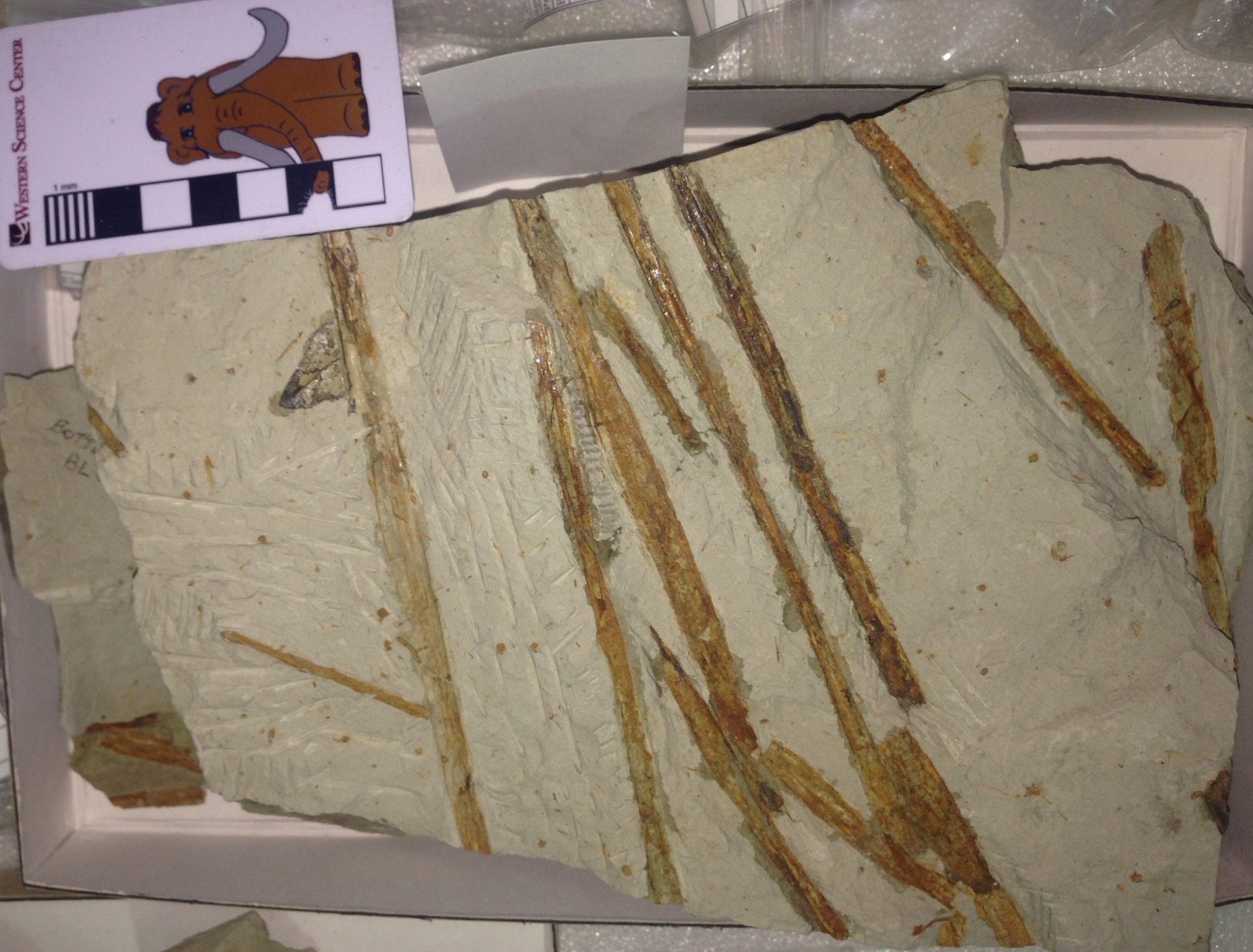 Fossil plants often don't get the attention they deserve, but besides being interesting organisms in their on right, they are in many ways far better indicators of past environmental conditions than are animals.Excavation of the Early- to Middle- Pleistocene deposits at the Southern California Edison El Casco Substation in San Timoteo Canyon produced numerous fossil plants, including quite a few examples of bur-reed (Sparganium sp.) such as the examples shown above. Sparganium is an aquatic plant that grows in marshes and standing water, and while several species still live in California we don't see them in areas as dry as San Timoteo Canyon is today. Several other marsh plants, including horsetails, were also found in these deposits.As I was photographing this specimen, I noticed another plant peaking out from under the bur-reed:
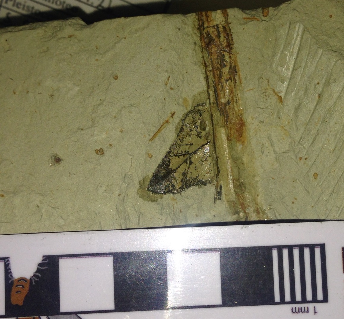 I'm pretty sure this is a birch leaf (Betula sp.), which is known from other specimens in the El Casco collection. Betula doesn't require as much water as Sparganium, but it's still not tolerant of arid conditions. However, today Betula is not found naturally anywhere in southern California, and in fact is found nowhere in the southern United States (even in humid areas) except at high elevations. It seems that birch are not tolerant of frequent high temperatures, and thrive in the cooler conditions found today at higher latitudes and altitudes. The presence of birch and abundant water plants such as Sparganium in the El Casco collection suggested wetter and cooler conditions at San Timoteo Canyon during the Early Pleistocene relative to what we see there today.
Fossil Friday - collared lizard vertebra
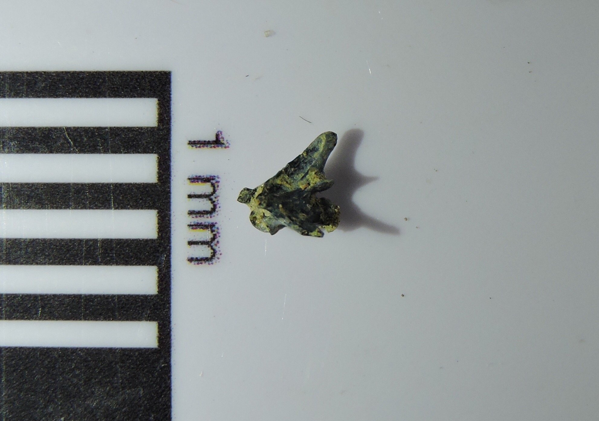 From a geological standpoint the Ice Age was not very long ago. Almost all the plant and animal species around today are survivors of the extinction that took place at the end of the Pleistocene. In many Ice Age deposits, while extinct megafauna get the most attention, the most widespread fossils are from species that are still with us today.Above is a caudal (tail) vertebra from a collared lizard of the genus Crotaphytus. It's shown in left lateral view, with the front of the bone to the left. Below is a dorsal view of the same bone, with anterior toward the top:
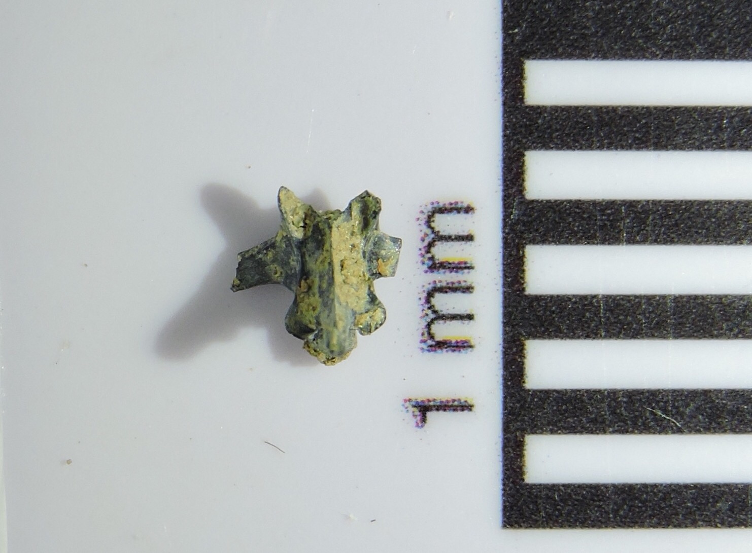 In this view we can see that the transverse processes that stick off each side of the vertebra are broken, as is the right prezygopophysis (the prezygopophyses are the bony spurs projecting off the front of the vertebra; they would articulate with the vertebra immediately ahead of this one). Besides this slight damage, the black color suggests this bone has been burned in a fire, as is the case with a number of other bones from the Diamond Valley Lake deposits.Collared lizards are widespread in Mexico and the southern United States west of the Mississippi River, especially in more arid environments. They are fast-moving carnivorous lizards that feed on insects and small mammals. There are several living species, some of which include brightly-colored males with banding around their necks that inspired their common name. Unfortunately, we probably can't identify the species of collared lizard based solely on an isolated tail vertebra, but we can say that these colorful lizards were present in the region just as they are today.
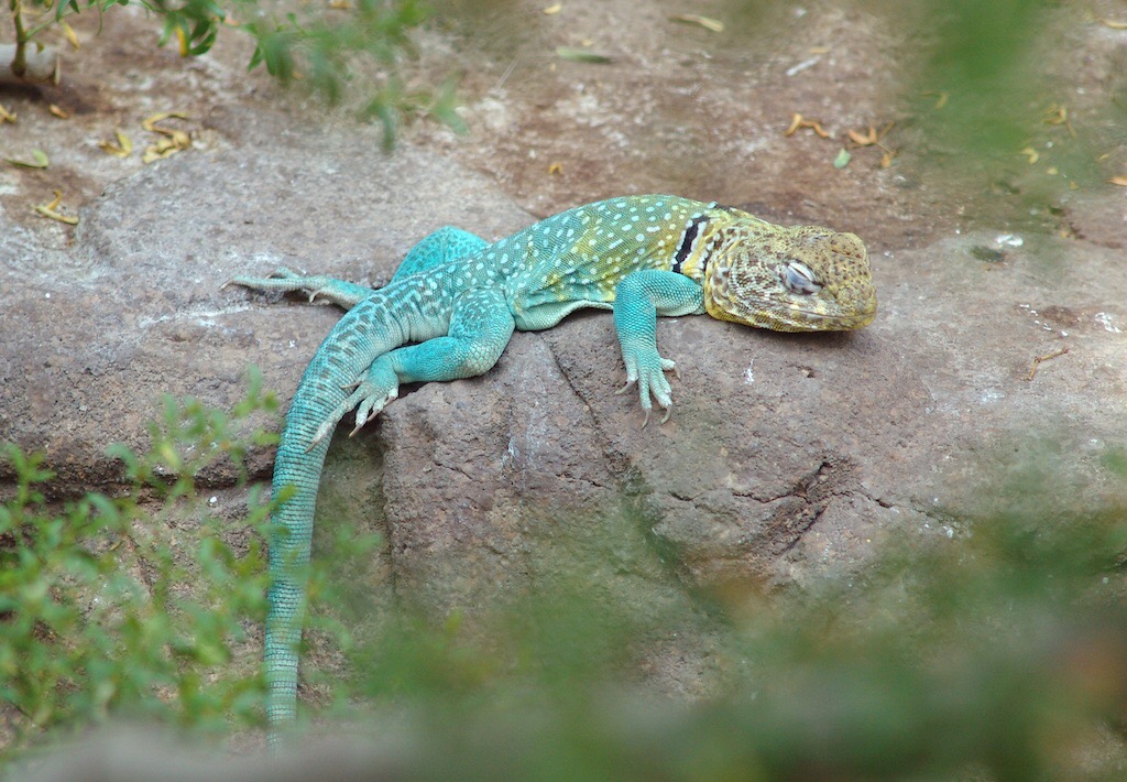
Fossil Friday - mastodon cervical vertebra
 Elephants and their relatives have big heads. They're big animals to begin with, but they also have strong, heavy jaws and teeth, as well as big brains. Then they throw in a massive trunk and huge heavy tusks (four of them, in the case of some extinct species). If you stick all these heavy features onto your skull, there are going to be consequences.Two things we very rarely see together in one animal are long necks and large heads. In some ways the head and neck act as a third-class lever, which actually increases the force necessary to move the head. The longer the neck the greater the necessary force. Significantly, that also applies to the force needed to stop the head from moving once it's started; without enough force, the head may keep moving independent of the neck and the rest of the body, with unfortunate consequences. What this boils down to is that animals with big, heavy heads will almost always have short necks.The image at the top is the third cervical (neck) vertebra from a mastodon, seen in anterior view, and below is the same vertebra in posterior view:
 The vertebra has both epiphyses attached, which suggests that it was not a particularly young animal (we can't say that it was fully mature, since the cervical epiphyses fuse at a relatively young age). There are some hints of osteoarthritis. This could indicate that the animal is older, but I wonder if this is common in elephant neck vertebrae, given all the stresses on the neck.The right lateral view is especially interesting:
 This vertebra is amazingly short. If I had found it in a marine deposit I would have probably initially thought it came from a whale - another large-headed animal with short neck vertebrae.In mastodons and other proboscideans, the third through seventh cervical vertebrae are all short like this. A string of these short vertebrae is not going to be very flexible, so one of the trade-offs in having a big, heavy head is having a short, inflexible neck. In most whales and dolphins some of the neck vertebrae are actually fused together making them completely immobile. This hasn't happened in elephants, but they still have very limited neck mobility. As an example, check out videos such as this one in which elephants are facing off with possible threats. Notice that as the elephant in the video looks in different directions, he's actually turning his whole body. His head is staying in almost the same position relative to his back the whole time. Elephants can still nod their heads and rotate their heads around the neck, because these motions are largely controlled by the first two neck vertebrae. But they have very little ability to point their head in a different direction than the rest of the vertebral column, a limitation that they share with their mammoth and mastodon relatives.
Fossil Friday - bison humerus
 At first glance, bison seem to be rather oddly proportioned, with a relatively massive head and shoulders. And sometimes, things are exactly as they appear; bison really do have huge, heavy heads. Carrying around such a large head has effects on the rest of the body, and bison have strong neck vertebrae, long neural spines on the vertebrae over the shoulders, and robust forelimbs to help support the weight of the skull. This was even more of an issue for Pleistocene bison such as Bison latifrons and B. antiquus, with their relatively larger and heavier horns compared to the modern B. bison.Today's Fossil Friday specimen is part of the right humerus (upper arm bone) from a Pleistocene bison:
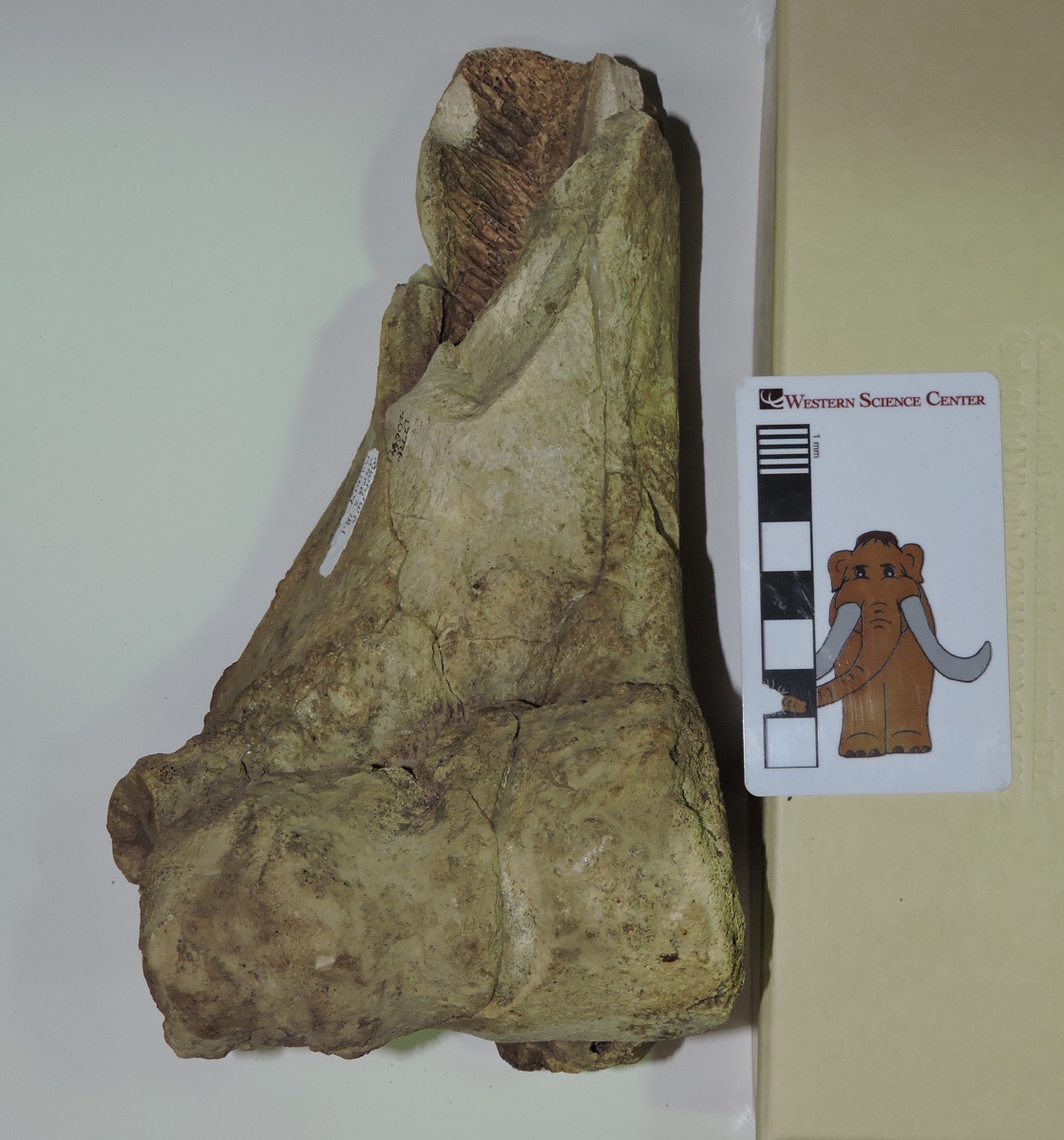 It's shown above from the front (anterior or cranial view). This is only the distal part of the bone, with the elbow joint at the bottom. The proximal half with the ball joint at the shoulder is missing. This is a big, heavy bone; the articulation surface at the elbow is about 9 cm across.Here's the same bone from behind (posterior or caudal view):
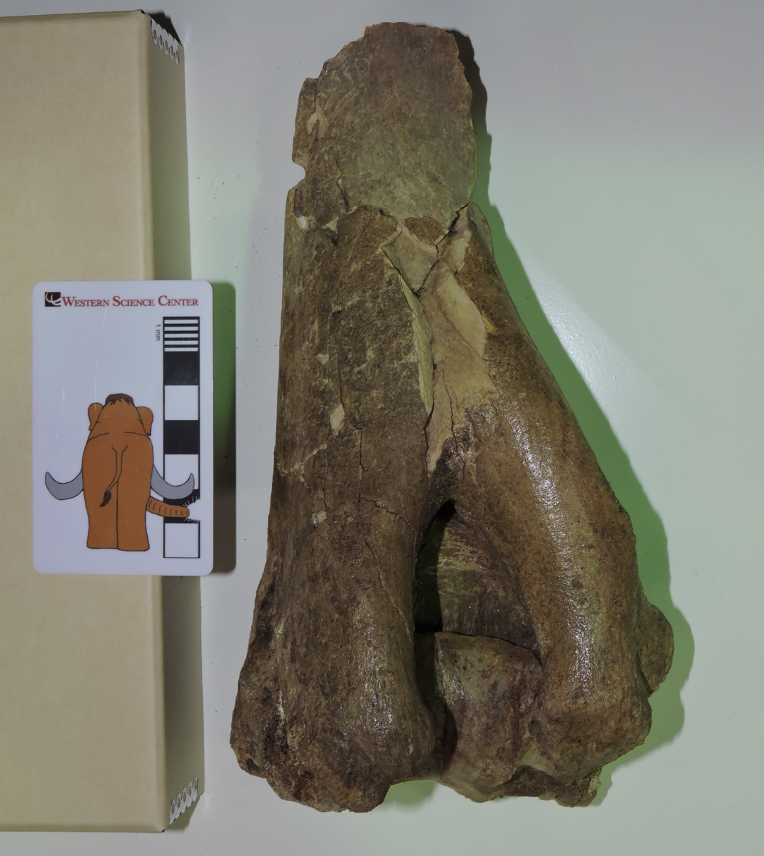 The large notch at the bottom is called the olecranon fossa. It accommodates a large projection on the connecting bone, the ulna, called the olecranon process. This process forms the point of the elbow, and serves as the attachment point for the triceps brachii muscle that extends the forelimb. When the triceps brachii is flexed, it straightens the forelimb, rotating ulna's olecranon process into the humerus' olecranon fossa.
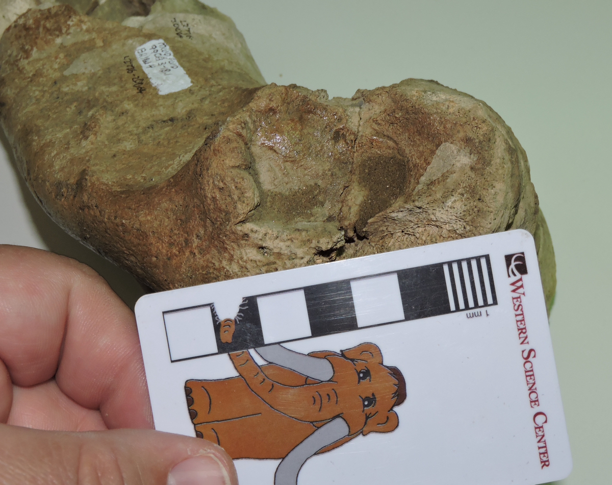 Above is an oblique view of the lateral side of the humerus. The circular area above the scale bar is called the lateral epicondyle, and in this specimen it has a somewhat irregular shape and texture. This could be due to osteoarthritis, which wouldn't be surprising in the elbow joint of a heavy-headed animal such as a bison. The distal end of this humerus is fully fused to the rest of the bone (unlike the mastodon humerus we featured last week), suggesting that this bison was full grown at the time of its death.
Fossil Friday - juvenile mastodon humerus
 It's been over two months since we've featured a mastodon for Fossil Friday, which seems a little odd for the Valley of the Mastodons, so this week we have a mastodon humerus.This is a left humerus (the upper arm bone), seen above in approximately anterior view. The proximal end (closest to the shoulder) is on the left, while the distal end (closest to the elbow) is on the right. Below is the same bone in posterior view:
 The ends of the bone are indistinct and have no obvious articulations with other bones. While there is some damage to the bone, the primary reason for this is that this was a very young mastodon. The humerus starts out as three different bony components, a main shaft and epiphyses at each end that are all held together by cartilage. As the animal grows the elements eventually fuse together into a single unit, but if the animal dies before it's fully grown the epiphyses may fall off as the cartilage decays. That's what's happened in this case, with both the missing epiphyses and the small size indicating that this was a very young mastodon.
Fossil Friday - Smilodon metacarpal
 Part of the nature of paleontology is attempting to pull as much information as possible out of often very limited data. As I mentioned last week, many fossil specimens may not be especially visually compelling, but nevertheless can provide useful information.A few weeks ago I discussed a bighorn sheep bone from a small collection of fossils recovered from Bureau of Land Management property near Desert Center in Riverside County. A number of other species were found at this site, with one of the most surprising being the sabertooth cat Smilodon.The only identifiable fragment from Smilodon that was found was the partial left second metacarpal (the hand bone that supports the index finger) shown above. It happens that at WSC we have a cast skeleton of Smilodon that we use for programming that allows us to place it in context:
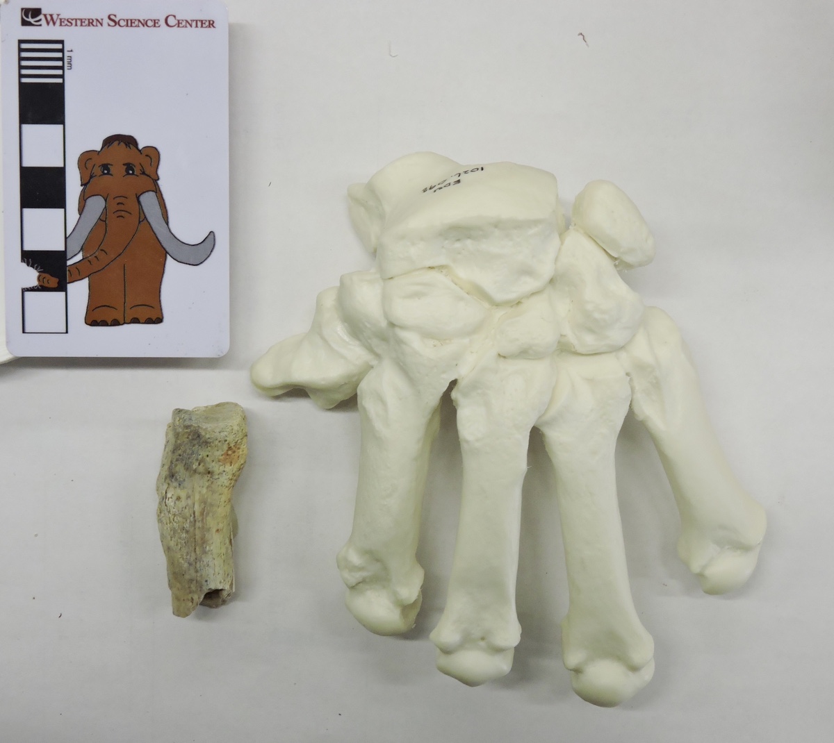 Here's a marked-up version, with the approximate preserved portion of the metacarpal indicated:
 So all we have is the proximal half of the bone, and even that is damaged. Nevertheless, the size and shape make this a pretty solid identification. The interesting thing is that this is the only reported Smilodon bone from the entire region around Desert Center, and in fact is the only record of any large Pleistocene cat from this area (Raum et al. 2014). So this tiny fragment turns out to be a relatively significant specimen for what it can potentially tell us about the range of Smilodon and the Pleistocene ecosystem in the Desert Center region.Reference: Raum, J., Aron, G. L., and Reynolds, R. E., 2014. Vertebrate fossils from Desert Center, Chuckwalla Valley, California. In R. E. Reynolds (Ed.), Not a Drop Left to Drink, California State University Desert Studies Center 2014 Desert Symposium: 68-70.
Fossil Friday - Stories from Bones exhibit
![]() For Fossil Friday this week, I want to highlight Western Science Center's new exhibit "Stories from Bones", which opens tomorrow.While WSC has excellent paleontology exhibits, as with any museum with a large collection many of the specimens are not on public display. There are a variety of reasons for this. Of course, the biggest obstacle is money; cases, information panels, interactive, floor space, and other requirements for an effective display are all expensive, and even the healthiest museums operate on a shoestring budget. Besides money issues, many specimens are just not suitable for display. Perhaps they're too fragile to risk moving around too much, or too fragmentary to interpret for the public (a specimen that visually looks like a piece of junk can still produce valuable scientific data). Even with all these limitations, we strive to make as much of our collections accessible to the public as possible. "Stories from Bones" is a result of that effort.
For Fossil Friday this week, I want to highlight Western Science Center's new exhibit "Stories from Bones", which opens tomorrow.While WSC has excellent paleontology exhibits, as with any museum with a large collection many of the specimens are not on public display. There are a variety of reasons for this. Of course, the biggest obstacle is money; cases, information panels, interactive, floor space, and other requirements for an effective display are all expensive, and even the healthiest museums operate on a shoestring budget. Besides money issues, many specimens are just not suitable for display. Perhaps they're too fragile to risk moving around too much, or too fragmentary to interpret for the public (a specimen that visually looks like a piece of junk can still produce valuable scientific data). Even with all these limitations, we strive to make as much of our collections accessible to the public as possible. "Stories from Bones" is a result of that effort.
 Mammoth jaw display in "Stories from Bones".
An important aspect of planning an effective exhibit is developing a theme. An exhibit is telling a story, and you need to be aware of what that story is as the exhibit is being designed. The theme might be "We have a bunch of stuff!", but while that was a common theme in museums a century ago (and one I personally appreciate), it does not generally make for the most informative exhibit experience for the majority of visitors.Once the theme is established, it's important to stick to it, so that the exhibit story remains coherent. Imagine reading a mystery novel in which three chapters are devoted to a history of the development of the gunpowder used in the crime, and an additional chapter describes the etymology of the last name of the victim, when neither is important to the outcome of the story. Each of these things might be individually interesting, but if you try to talk about all of them then you risk obscuring everything. There is a real risk of this "mission creep" in an exhibit based on a data-rich field such as paleontology. We might talk about evolutionary relationships, paleoenvironmental indicators, biogeographic information, site-specific descriptions, or an array of other things. Talking about any of these might be a good idea; talking about all of them is a bad idea.The permanent paleontology exhibit at WSC does this very well. The exhibit is basically a review of the Diamond Valley Lake Local Fauna; what was here, how does it compare to the rest of Southern California, and (as a secondary point) what does it tell us about the local Pleistocene paleoenvironment. In contrast, "Stories from Bones" asks "What do these fossils tell us about the lives and deaths of these individual animals?".To that end, "Stories" has a series of displays that talk about how paleontologists determine how old an animal was when it died. We have several cases that look at tooth replacement in proboscideans, horses, and bison, such as the two mammoth jaws above (they're close to the same size, but one animal was about 30 years older than the other), or the three bison dentaries shown below that represent young, middle-aged, and elderly animals.
 Bison jaw display in "Stories from Bones".
We have several examples of bones that were broken and healed, evidence of events that took place during an animal's life:
 Broken and healed bones in "Stories from Bones".
We also have several cases that describe taphonomic features, looking at what happened to an animal at or immediately after death.We designed and built a number of interactive displays for this exhibit. The most prominent is a cast and video of the CT scans of Max the Mastodon's lower jaw, taken back in August.
 Max's CT-scan station in "Stories from Bones" during installation, under the watchful eye of @MaxMastodon.
We're proud of the fact that several of our interactive displays ask visitors to map or measure specimens and reach conclusions based on their data:
 A more extensive version of the bison tooth display shown here is also available as a guided activity for school groups visiting the museum, and as a kit available for purchase.If you're a regular reader of this blog, you'll find that "Stories from Bones" draws heavily from my past "Fossil Friday" posts. For most of those specimens, this is the first time they've ever been on public display, so if you're near Southern California make sure to stop by the museum. "Stories from Bones" opens on October 31, and will remain open into May 2016.
