

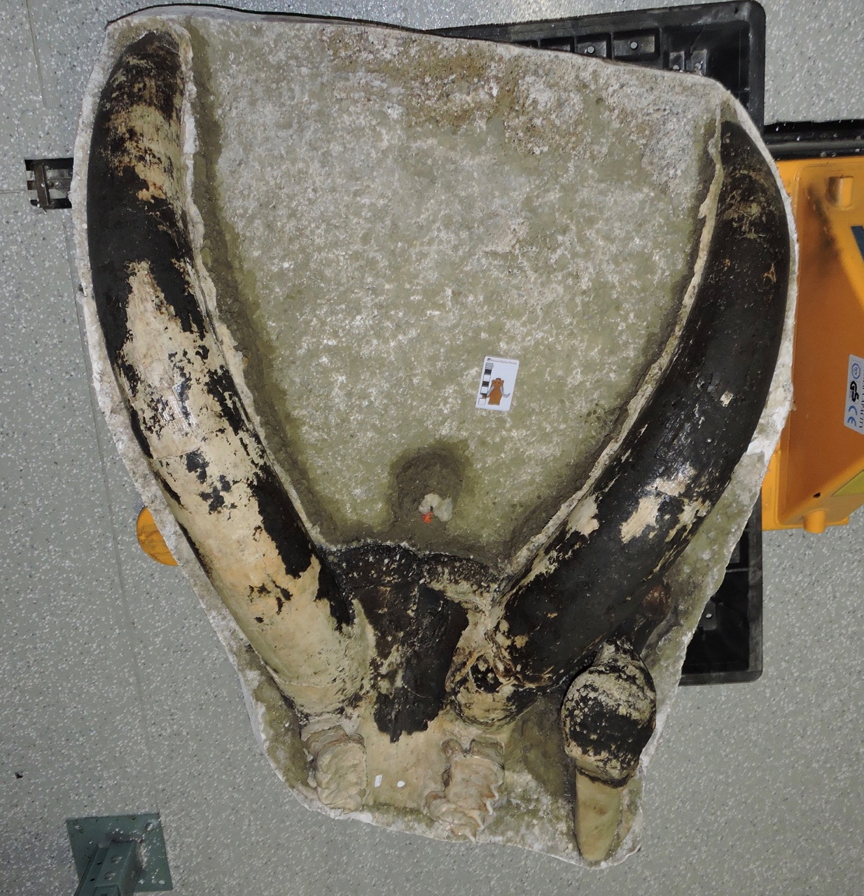
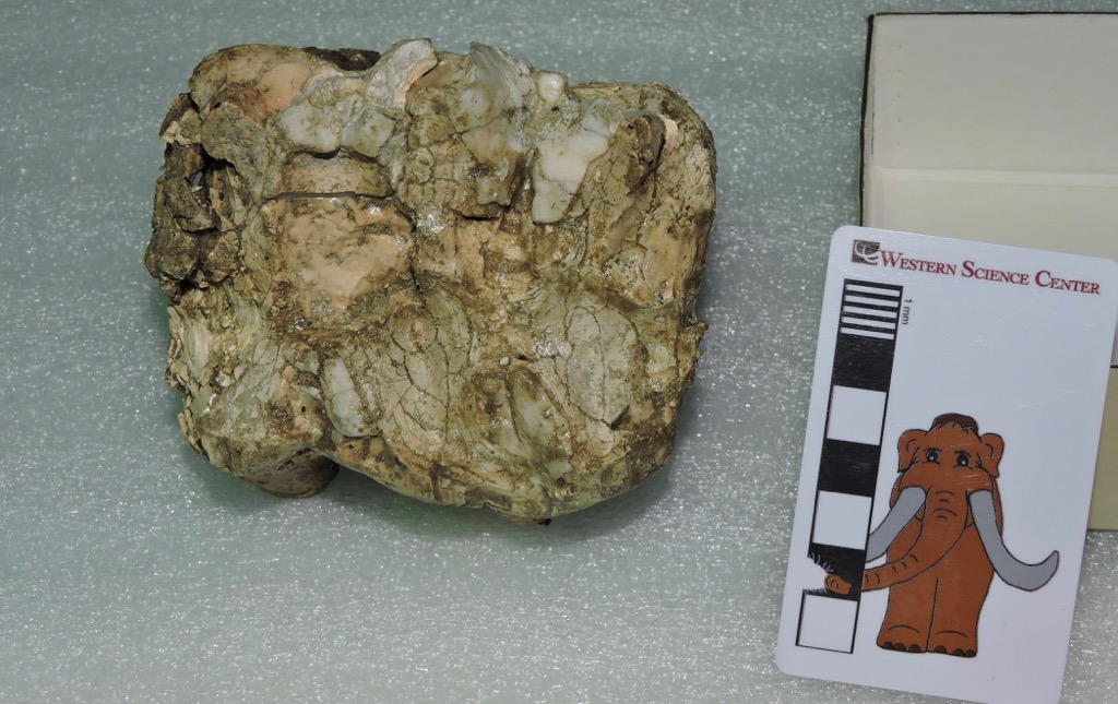
 Today is World Elephant Day, recognizing the conservation difficulties faced by the surviving species of elephants. Last week, with Katy Smith's visit to WSC to examine mastodons and Bernard Means' visit to 3D-scan some of our specimens, as well as needing more data for the Mastodons of Unusual Size Project, we had the opportunity and motivation to open a lot of mastodon jackets that have remained unexamined for years. This confluence of events make an excellent excuse for featuring another mastodon for today's Fossil Friday.It turns out that, as much as I've touted the mastodon collection at WSC, it's even better than I had realized. Shown above is one of several partial skulls we examined last week. The skull is oriented ventral up, so the palate side is visible; the lower jaw is not preserved. Anterior is to the top of the image. The two large tusks are essentially complete, as is the ventral side of the premaxillary bone that supports them. A small portion of the maxilla is preserved, which includes both upper second molars and the anterior portions of the third molars, which are more clear in the image below:
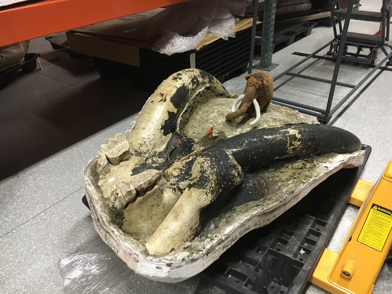 The proximal end of one femur is also preserved; it's the lump of bone in the foreground.The most notable thing about this skull is the black coloring over much of the surface, including almost all of the left tusk. It appears that this specimen was burned, almost certainly after death. We see this in a fair number of bones from Diamond Valley Lake, but it's the first time we've noted it in a mastodon, leading our Marketing Specialist Brittney Stoneburg to nickname this specimen "Blaze".Blaze is missing most of his third molars, but his second molars are intact and we were able to get measurements for the Mastodons of Unusual Size Project. Katy was also able to get measurements for her studies on mastodon tusks (and, based on the size of the tusks, Blaze was almost certainly a male). Blaze's second molars were heavily worn, and the anterior portions of his third molars were in wear, suggesting that he was close to the same age as Max when he died, perhaps in his mid-30s.Today on World Elephant Day, help support elephant conservation by visiting or donating to a museum, zoo, or conservation group that furthers our knowledge and protection of these amazing animals.
Fossil Friday - associated mastodon material
 This week we have two visiting researchers at Western Science Center. Dr. Katy Smith from Georgia Southern University has been measuring and photographing the proboscidean tusks in our collection, which we hope will lead to all kinds of new information about southern California mastodons and mammoths. Dr. Bernard Means from Virginia Commonwealth University's Virtual Curation Lab has been here on a trip sponsored by Smithsonian Affiliations to make 3D scans of some of the WSC specimens (Bernard has written about his visit here). These visits have meant that we've been pulling out lots of specimens, many of which I had never seen before.Last June I wrote about one of our mastodon jaws, which is an interesting specimen for the Mastodons of Unusual Size project because of its outlier status. This mastodon has the widest lower third molars we've measured in any California specimen, by a pretty large margin. As it turns out (and unknown to me until this week), we have part of the cranium from the same specimen.In the image above, the cranium fragment is sitting beside the lower jaw, and facing the opposite direction, so anterior is to the left. The fragment consists mostly of the left premaxilla and maxilla with the associated teeth. The rather large left tusk suggests that this was a male mastodon (the distal end of the tusk is broken off). The left second and third molars are both well preserved, with the second molar fully in wear and the third molar just starting to erupt, suggesting he was in his early 20's (and consistent with the lower jaw tooth wear).It turns out the upper third molars are outliers, but not quite to the same degree as the lower molars. We have several California upper M3s that are wider than this tooth, but those teeth are also much longer than this one. That means that the proportions of this tooth are unusual for California, even though we have teeth that are both longer and wider than this one. In fact, there's a tooth in our database from Ohio that is almost exactly the same dimensions as this one.
Fossil Friday - Isorthoceras sociale
 During the Mastodons of Unusual Size road trip, Brett and I made several stops to collect fossils for WSC. Since I was 20 years old, I've made several visits to the small, well-known (to paleontologists) roadcut in Graf, Iowa (the photo below was from a trip I made there in 2007):
 The rocks at Graf are from the Late Ordovician Maquoketa Formation, about 445 million years old. A number of different types of marine invertebrate fossils are found here, but perhaps the most prominent is the cephalopod Isorthoceras sociale.Cephalopoda is the group of mollusks that includes modern squid, octopus, and cuttlefish. Another group of modern cephalopods are the nautiloids, such as the chambered nautilus:
 Unlike other living cephalopods, nautiloids have an external shell made of the mineral aragonite. The shell in modern nautiloids is shaped like a coiled cone, and is divided internally into chambers:
 While nautiloids are relatively uncommon today, for hundreds of millions of years they were among the most abundant animals in the ocean, and it was during the Ordovician Period that they first became widespread. While the living nautilus has a coiled shell, nautiloid shell forms were more diverse in the past. Many of the early nautiloids had straight, conical shells. At Graf, there are several rock layers that are made up almost entirely of the shells of the small straight-shelled nautiloid Isorthoceras, at least four of which are visible in the photo at the top of the page. Here are some reconstructions of what Isorthoceras may have looked like:
 Most of the shells in any given layer at Graf are aligned in the same direction, so from certain angles the shells are mostly seen in cross section:
 The shells also sometime erode out of the rock, and can be found as isolated fossils:
 During our short stop at Graf, Brett and I filled a 5-gallon bucket with rocks containing Isorthoceras and other Ordovician marine fossils which will be added to the WSC collections.
Fossil Friday - mastodon astragalus
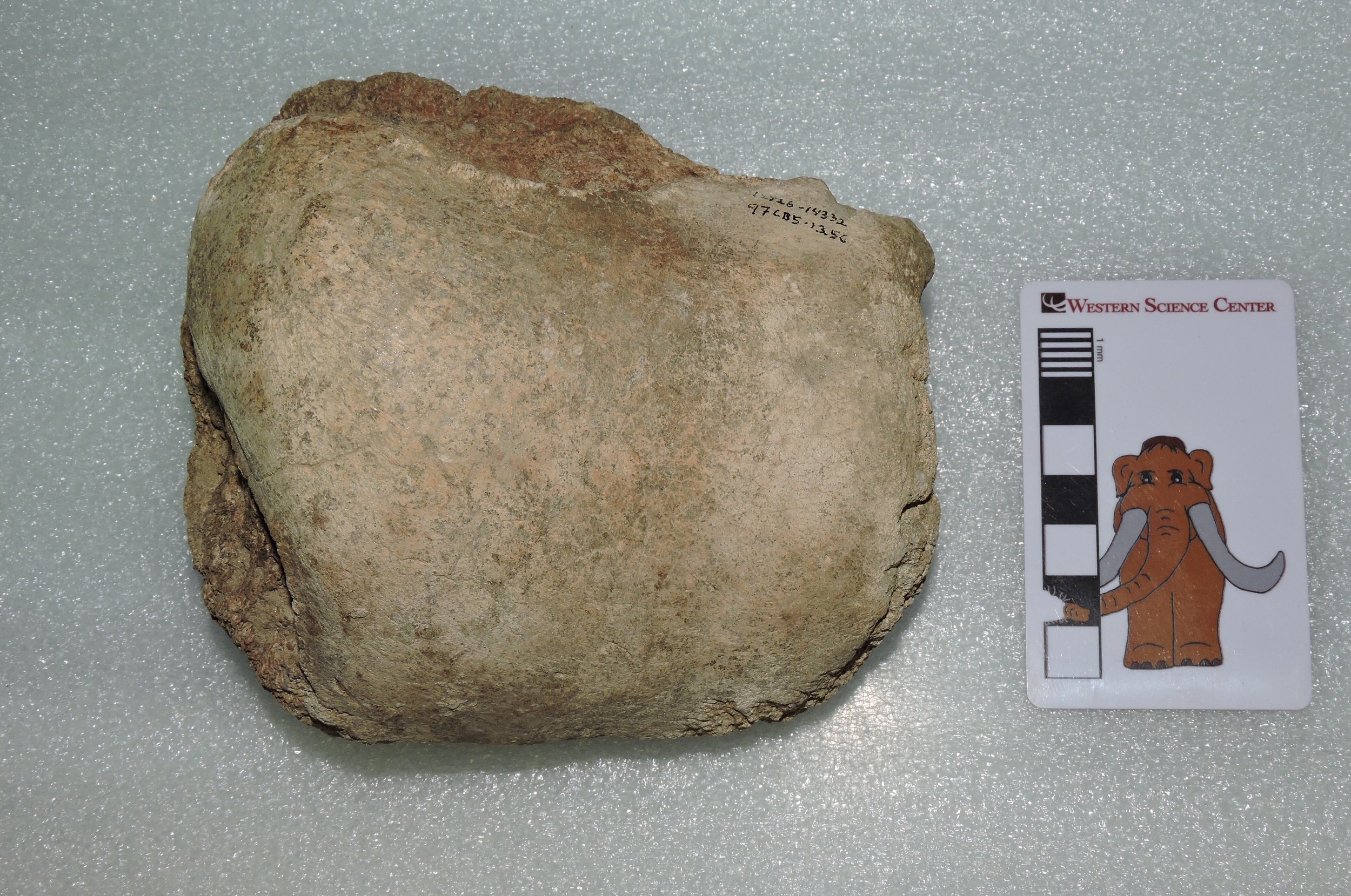 I'm still on the experiment.com-funded Mastodons of Unusual Size tour, and will be doing an additional post on that tomorrow. My inability to type and drive at the same time means I can only do a short Fossil Friday post.The bone shown above is a mastodon astragalus, the most proximal of the ankle bones. The upper end of the astragalus articulates with the lower ends of the shin bones (the tibia and fibula), although in elephants the amount of movement at that joint is limited. Proboscidean ankle bones are much less complex than in most other animals, and are optimized for weight-bearing. Interestingly, it seems that some dwarfed elephants evolved a rather different ankle structure for walking in hilly terrain.This astragalus was found at the East Dam of Diamond Valley Lake.
Fossil Friday - mastodon thoracic vertebra
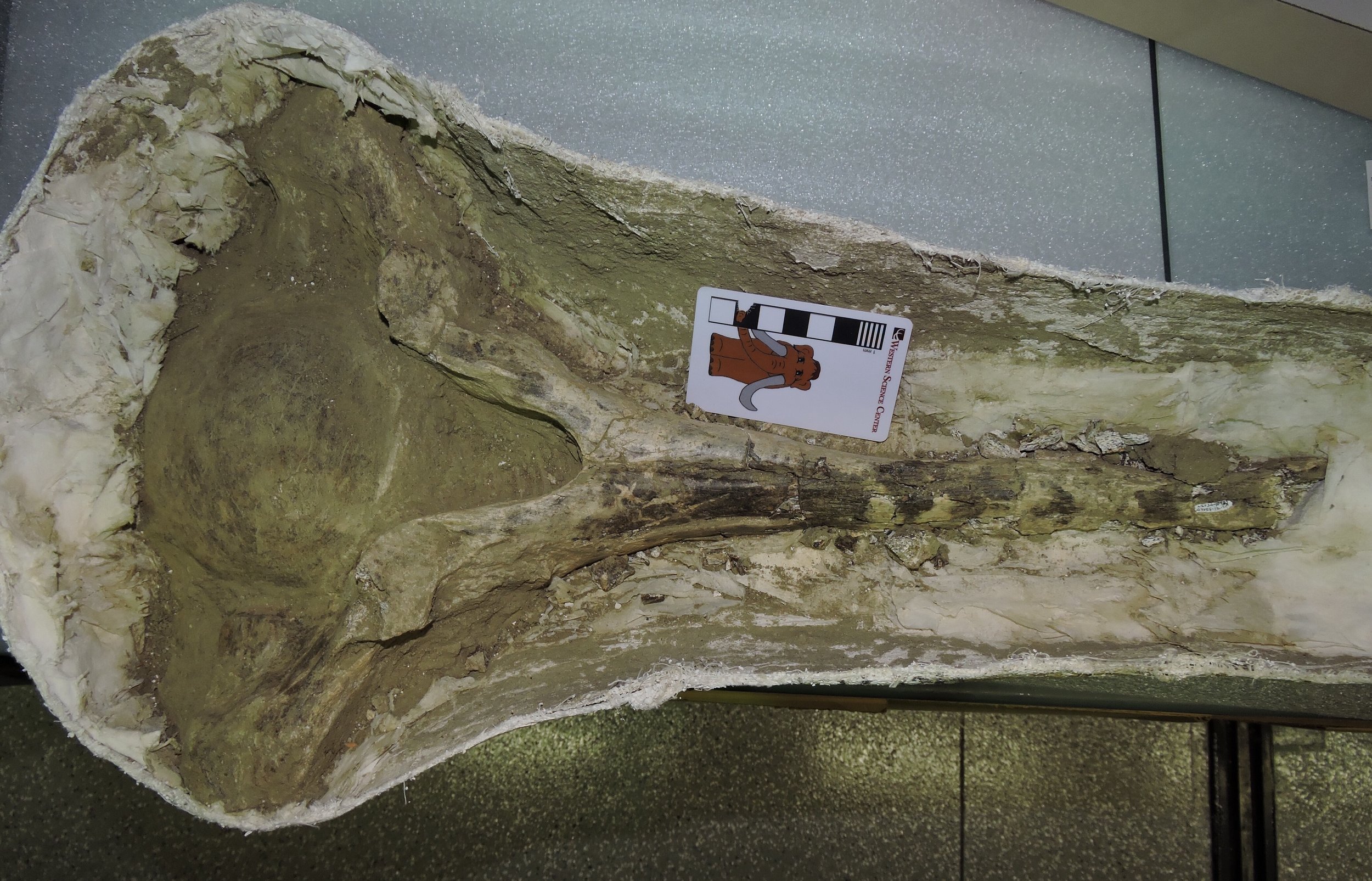 As I'm in the middle of our Mastodons of Unusual Size data-collecting trip, my Fossil Friday post will have to be a short one. But, in keeping with our road trip topic, it will of course feature a mastodon!The specimen shown above is an anterior thoracic vertebra, shown in anterior view. The image is turned sideways to fit better on the screen, so the bottom of the vertebra is on the left. The bone is still in its field jacket, and has only been partially prepared.Thoracic vertebra in mammals are the ones that articulate with the ribs, and the articulations are visible on the ends of the processes projecting from each side of the vertebra. The tall neural spine extends from the top of the vertebra (right in the photo). The neural spines serve in part as muscle attachment points for muscles that hold up the head and help support the front legs. In quadrupedal animals with large, heavy heads, such as mastodons, the anterior neural spines are very long, providing more surface area and better leverage for those muscles.This vertebra was collected from the Diamond Valley Lake East Dam, close to where the Western Science Center is now located.
Fossil Friday - mastodon molar
 Sometimes the fossils we recover are a little worse for wear. The remains may have deteriorated before burial, been damaged or deformed by sediment weight, structural forces, or groundwater after burial, weather out of the sediment and get eroded before discovery, or damaged during the act of discovery or excavation (I've collected numerous fossils that were "discovered" by bulldozers!). Sometimes it seems amazing that any fossils reach us at all. And yet, even these damaged specimens can be significant.The rather amorphous lump above is actually a mastodon tooth from Diamond Valley Lake, seen in occlusal view (the chewing surface). If you don't look at mastodon teeth on a regular basis, it might be a little difficult to recognize at first, because a lot of the enamel is missing. It's much more recognizable in lateral view, although the damage to the crown is still obvious:
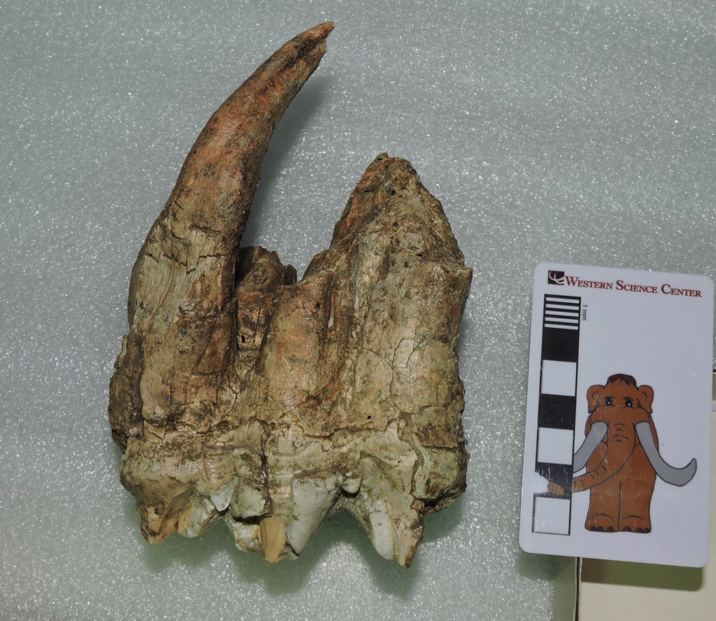 Even with the imperfect preservation, there's quite a lot we can say about this tooth. The lophs (the enamel ridges on the crown) are nearly perpendicular to the long axis of the tooth, suggesting that this is an upper rather than a lower tooth. The curvature of the roots indicates that the anterior side of the tooth is on the left in the photo above. Upper mastodon teeth wear more rapidly along the inside edge (the lingual side) than along the outside edge (the labial side), so that makes this an upper left tooth. There are three lophs present so that limits the tooth to either the fourth premolar, the first molar, or the second molar (the second and third premolars only have two lophs each, while the third molar has four or sometimes five lophs). From P4 to M2 the teeth get progressively larger, and this tooth is pretty large. Combining all these observations, the tooth is the upper left second molar.But wait, there's more! Even though a lot of the enamel is missing, there's enough to tell that the tooth was fairly heavily worn, so it had erupted before this animal died. But the crown wasn't completely worn away; there are still grooves separating the lophs, at least on the labial side, so this tooth was probably still in use when the animal died. For example, Max's 2nd molars, which are still in his jaws, are more worn than this one. That means the mastodon was a young adult when it died. This is supported by the long, intact root, indicating that the tooth had not been shed. Instead, this tooth was still in the mastodon's skull when the animal died, and the skull was broken up at some point, probably before burial.Finally, there is actually enough of this tooth preserved that we will be able to include this specimen in our study of mastodon tooth proportions, since we can get good crown length and width measurements.Brett and I leave this morning on our museum tour to collect measurements on mastodon teeth from other areas, visiting at least five museums across the central U. S. during the next two weeks. I'll be posting updates about our trip on this blog and on experiment.com/mastodons and Twitter (@AltonDooley), as well as on the museum's Instagram and Facebook pages. Max's alter ego is also accompanying us, and will be posting his own updates on his Twitter feed (@MaxMastodon).
Fossil Friday - mastodon mandible
 In a week Brett and I head out on our experiment.com-funded road trip to collect data on mastodon teeth. The major goal of this project is to collect enough data to better understand what's going on with mastodon tooth proportions, especially with respect to the apparently unusual California mastodons.The nicely preserved lower jaw shown above is a WSC specimen recovered from the San Diego Canal near Diamond Valley Lake, and which was included in our recently-closed "Stories from Bones" exhibit. Below is a dorsal view:
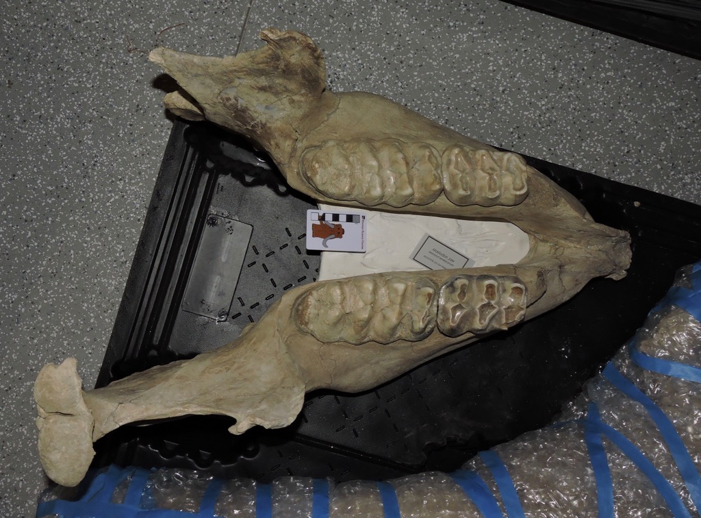 This animal was a little smaller than Max, and based on tooth wear, a bit younger. While the first molars are lost, the third molars are just coming into wear, so this animal was likely in its early 20's.This specimen is of particular interest in our project, because it's an outlier. So far, this mastodon has the widest lower third molars of any California mastodon we've measured, and it's not really even close. The left molar on this specimen is almost 89 millimeters wide, while the next largest California specimen is just over 83 mm (the right molar is a bit narrower, but is still almost 85 mm). This is the only California specimen we've seen that has m3 proportions that approach typical Florida or Indiana specimens.The outlier status of this specimen is even more noticeable because, of the 14 California mastodons with m3 measurements currently in our database, 5 have m3 widths between 82.9 and 83.1 mm. That width seems to be a sharp line that California mastodons almost never cross, with the exception of this one specimen. (As an aside, gigantic Max's m3 is only 76.6 mm wide, one of the narrowest in our data set).As we examine additional California specimens, it's likely we'll eventually find other mastodons with teeth that are comparable to this specimen. But for now it stands out as the only California mastodon that has tooth proportions that approach those of eastern specimens.
Fossil Friday - horned lizard spike
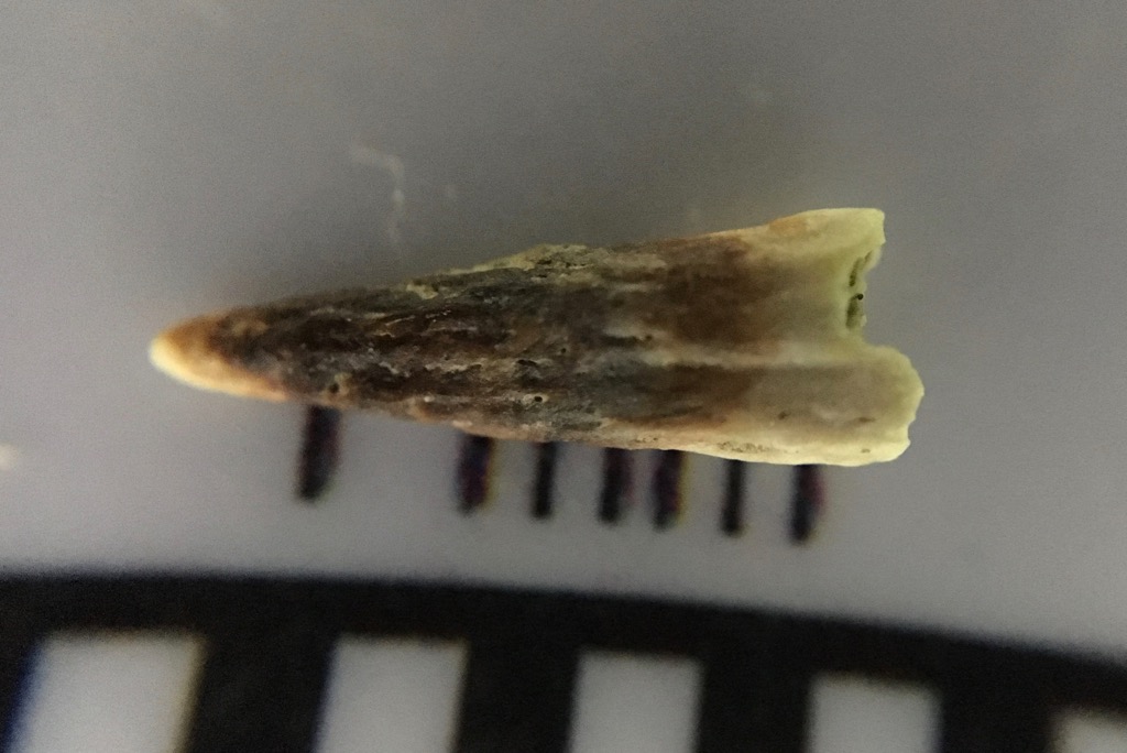 One of the things I find most intriguing about the Pleistocene is how, in spite of the impressive megafauna, it was so similar to the modern world. As I've mentioned in other posts, as with many other Pleistocene sites the fauna at Diamond Valley Lake is in many ways a lot like what we see in southern California today.The tiny fossil shown above (that's a millimeter scale) may look like a tooth at first glance. But the pitted texture is a giveaway that this is a bone, not a tooth. This is a horn from a horned lizard of the genus Phrynosoma, specifically one of the horns that projects from the parietal bone in the skull, visible below in this specimen of Phrynosoma cornutum that Brett photographed in Texas:
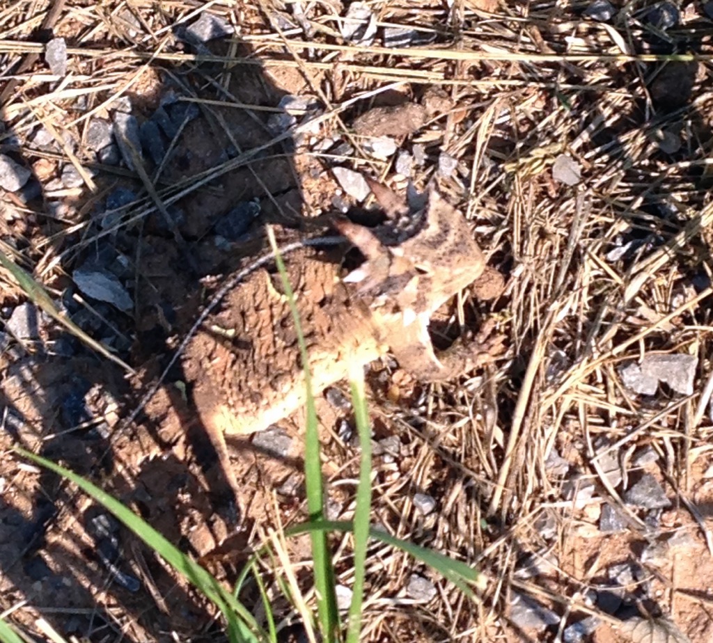 There are a number of different species of horned lizards that are each endemic to different parts of North America, and one of the ways they differ is in the size and shape of their horns. Compare the horns on the Texas lizard above to those on Phyrnosoma douglasii from South Dakota:
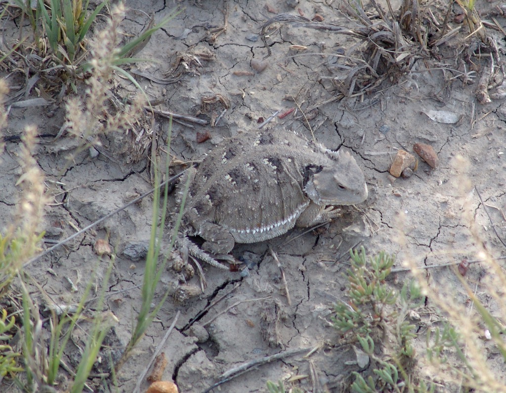 The southern California horned lizard species is Phrynosoma coronatum, which I've never had the opportunity to photograph in the wild. Its skull, however, is somewhat similar to the Texas species with large parietal horns, as can be seen in this comparison image.Horned lizards live in relatively arid areas, which seems to be a common theme with the small Pleistocene animals found at Diamond Valley Lake. They are specialist predators of ants, especially harvester ants, so the presence of Phyrnosoma at Diamond Valley Lake implies the presence of ants there as well, even though they have not been found as fossils in those deposits.
Fossil Friday - Max's femur
 An oddity in the preservation of Max the mastodon is that, while the skull and pelvis were beautifully preserved, very little was recovered from the massive limb bones. The only significant piece was the distal end of the left femur, and a few months ago while working on our mastodon project we opened our exhibit case to photograph and measure this fragment.The image above is in anterior view. The smooth curved surface at the bottom of the bone is part of the articulation for the knee joint. Below is the same fragment in posterior view:
 One of the reasons we wanted to get measurements of this bone is that we've often stated (following Springer et al., 2009) that Max is one of the biggest mastodons known from the western United States. The femur is one of the better bones for estimating body size, because in most terrestrial animals it's the primary weight-bearing bone, so its dimensions are often indicative of both height and weight. Ideally, we would like to have the entire femur to measure, but we take what we can get. In Max's case we can get a good measurement of the maximum distal width, which gives us a point of comparison to other mastodons. The graph below shows how Max's femoral width compares to mastodons from Rancho La Brea and New York:
 The two left bars are Rancho La Brea specimens, the tall bar in the middle is Max, and the two bars on the right are respectively the Watkins Glen mastodon (an adult male) and North Java mastodon (an adult female) from New York (measurements from Hodgson et al., 2008). Max's femur is quite wide compared to other mastodons, which was part of the reason Springer et al. suggested that Max was such a large animal.Using these measurements, I made a sketch estimating the total size of Max's femur and compared it to images of the same four specimens from the graph, with the caveat that proportions often don't scale exactly linearly so this is only an approximation. In addition, since Max's femur is the left one and the other four specimens are right femora, I've reversed Max's image so they're more directly comparable. (The Watkins Glen and North Java images are from Hodgson et al., 2008):
 Any way you look at him, Max seems to have been a pretty big mastodon. Yet Max's teeth are relatively small. This disparity in Max's measurements is what started us looking at the tooth shape and size in California mastodons, and is something I'll be exploring further next month as part of our mastodon project.References:Hodgson, J., W. D. Allmon, P. L. Nester, J. M. Sherpa, and J. J. Chiment, 2008. Comparative osteology of Late Pleistocene mammoth and mastodon remains from the Watkins Glen Site, Chemung County, New York. In W. D. Allmon and P. L. Nester, eds., Mastodon Paleobiology, Taphonomy, and Paleoenvironment in the Late Pleistocene of New York State: Studies on the Hyde Park, Chemung, and North Java Sites. Palaeontographica Americana 61:301-367.Springer, K., E. Scott, C. Sagebiel, and L. K. Murray, 2009. The Diamond Valley Lake Local Fauna: Late Pleistocene vertebrates from inland Southern California. In L. B. Albright, ed., Papers on Geology, Vertebrate Paleontology, and Biostratigraphy in Honor of Michael O. Woodburne. Museum of Northern Arizona Bulletin 65:217-235.
Fossil Friday-camel humerus
 In spite of the facts that camels are among the more common large animals from Diamond Valley Lake and are intrinsically cool, I've somehow managed to get almost halfway through 2016 without featuring them on Fossil Friday. I'll rectify that today.Above is a left humerus (upper arm) from Camelops hesternus. It's shown anterior (or cranial) view, with the proximal (upper) end on the left. The profusion at the lower left corner is part of the humeral head, the "ball" part of the "ball-and-socket" joint at the shoulder, while the articulation for the radius and ulna (the elbow joint) is on the right.This bone is still in its field jacket, and like many of the large bones from Diamond Valley Lake has only been partially prepared. Slowly but surely we're going to be finishing the preparation work on these bones, but it's a process that will take many years.
Fossil Friday-horse skull fragments
 Vertebrate paleontologists rarely get to examine whole skeletons (really, almost never). Even individual bones or regions such as skulls are usually fragments, often broken in odd ways, and identification can be a challenge. That's why paleontologists will keep around lots of well-illustrated references, reference collections of modern animal skeletons, and increasingly, 3D images of identified specimens. A lot of identification work is just comparing your unknown specimen to known ones and finding a match.For the last several weeks WSC volunteer Nathan Bonde has been preparing a block of sediment that came to the museum through a mitigation project (unfortunately with rather limited data). The sediment had several bone and possible tooth fragments sticking out, but it wasn't immediately clear what the material was or if it all belonged to the same animal.
 It quickly became apparent that the tooth was from a horse, apparently a upper left 3rd premolar or 1st molar (shown below next to a zebra skull from Brett's collection):
 The bone fragments were a little tougher. Continued cleaning showed that most of the bone joined together into a single large fragment, shown at the top of the page. The size and overall complexity suggested a skull fragment, which was bolstered by the presence of a bony tube that looked like the external auditory meatus, part of the ear structure. Since we knew that a horse was a strong possibility (based on the tooth), we compared the fragment to the zebra skull:
 If you're having trouble seeing how these match up, here's an annotated version:
 The area outlined in red on the zebra skull is the approximate preserved part of the fossil; note that the fossil is quite a bit larger than the modern specimen. The numbered features are:
- Right occipital condyle (the articulation with the 1st neck vertebra)
- Condyloid fossa
- Jugular process
- External auditory meatus
- Glenoid fossa (the articulation point for the lower jaw)
- Temporal fossa (opening for the muscles that close the lower jaw)
- Zygomatic process of the squamosal (cheekbone)
Here's another view, oblique and from behind, showing more clearly how the zygomatic process projects out, forming the lateral side of the temporal fossa: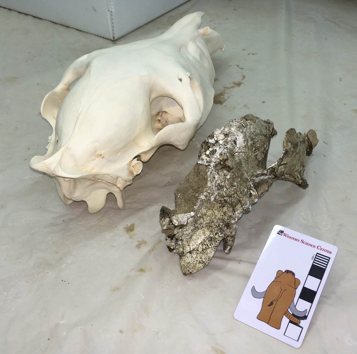 Tooth size is apparently not a reliable indicator of body size in horses, and our modern specimen is both sub-adult and a relatively small animal. Nevertheless, in this instance we seem to be dealing with a fairly large horse; both the tooth and the skull fragment are much larger than our reference skull. If you look closely at the images above, you may also notice differences in the shape of the enamel ridges on the teeth, and somewhat different shapes and orientations in some of the cranial bones. While both these skulls are from the genus Equus, they do not represent the same species, which isn't really surprising considering that they're separated by at least 10,000 kilometers and at least 20,000 years.
Fossil Friday-mastodon jaw fragments
 We're entering the final hours of our crowdfunding campaign at experiment.com, and are very close to our funding goal. This week we're featuring another specimen that will be included in this study.While the bulk of the Western Science Center's collection came from Diamond Valley Lake, we are a regional repository with a number of collections from other localities. This specimen was recovered during a mitigation project in Temecula in southwestern Riverside County, and after reconstruction by WSC volunteers it became apparent we had a significant portion of a mastodon lower jaw. The image is in dorsal view, with anterior to the left. Parts of both dentaries are preserved, although there is considerably more of the left dentary present.Portions of four teeth are preserved, the lower second and third molars. On the left side the second molar is complete, and the third molar is nearly so, allowing us to get length and width measurements for inclusion in our project; it turns out that these teeth are long and narrow, exactly what we've come to expect from California mastodons. The second molars are pretty heavily worn, while the third molars show wear only on the first two lophs. That is very close to the same wear state found in Max, so these two were probably close to the same age.If you haven't already done so, please go to experiment.com/mastodon and donate to our project, so we can start to understand how mastodons were distributed through North America.
Fossil Friday - Bison latifrons skull
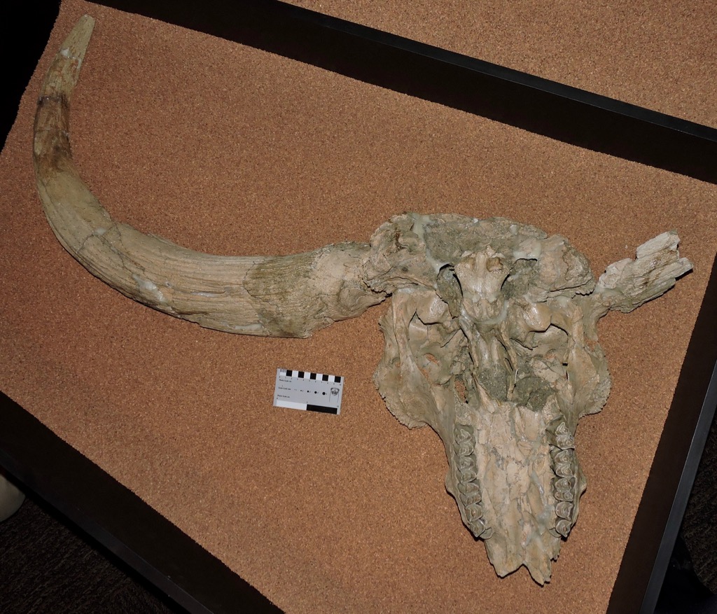 We still have a few days left in our crowdfunding campaign to study mastodons, but for Fossil Friday this week I'm stepping away from mastodons to look at a different animal. Earlier this week the U. S. Government passed a law designating the American bison (Bison bison) as the United States' National Mammal. Bison are prominent in the fossil record, including at Diamond Valley Lake.As I've mentioned in previous posts, there were two species of bison present at Diamond Valley Lake. The more common species was Bison antiquus, a possible direct ancestor to the modern B. bison. The rarer, larger species was the long-horned bison, B. latifrons.Diamond Valley Lake's best B. latifrons skull, shown above, is on permanent exhibit at the museum. It's displayed in ventral view, and in the image the front of the skull is at the bottom. The premaxillae forming the tip of the upper jaws is missing, as is the right horn core, but otherwise the skull is mostly complete if a bit deformed. One thing to keep in mind when looking at fossils of Bison latifrons (and, indeed, any fossil bovid) is that the preserved horn is simply the bone core of the horn. In life the core is covered with a keratin sheath, making the living horn considerably larger.It's somehow fitting that the United States' national mammal is in fact an immigrant to North America, and a relatively recent one at that. Bison were one of the last large mammals from the old world to spread across North America (some of the other large Old World immigrants, such as deer, mammoths, and mastodons, and South American ground sloths, all arrived earlier). In fact, bison only made it south of the 55th parallel (which runs through central Canada) roughly 250,000 years ago, and then rapidly spread across southern Canada and the United States. The first appearance of bison south of the 55th parallel is used by paleontologists at the marker for the start of the Rancholabrean North American Land Mammal Age (NALMA), the last NALMA of the Ice Age.During the Rancholabrean, various bison species ranged across the entire northern hemisphere. While some bison species went extinct at the end of the Ice Age, the genus Bison survived and thrived in the Holocene in both Eurasia and especially in North America. Two extant species are recognized, the American bison (Bison bison) (seen at Yellowstone National Park) and the European bison (Bison bonasus) (seen at the San Diego Zoo Safari Park):
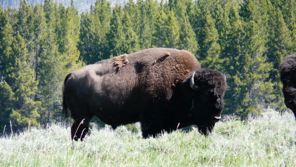
 This wasn't to last, as bison were almost completely wiped out by overhunting and habitat loss over the last few hundred years. Both species have been protected and are slowly recovering, although populations were reduced to such a low number in the past that genetic diversity is quite low. Even so, there are once again wild herds of both species living in protected areas.
Fossil Friday - Old Man Mastodon
 Closing our our Month of Mastodons is one of the more complete mastodon skulls from Diamond Valley Lake, a specimen that has several interesting features in addition to its relatively good preservation.First, to explain what you're looking at, here's an annotated version:
 The skull is laying on its right side, at a slight angle. Much of the braincase seems to be missing, although this area hasn't been fully prepared, so there could be more there than there appears. The left dentary (lower jaw) is present, but disarticulated; it's not clear if the right dentary is still in the bottom of the jacket.So, what can we say about this skull? Well, for starters it looks pretty large, with a massive tusk. I haven't measured it out yet, but I suspect that based on size this will probably be a male mastodon. Zooming in on the teeth gives us more information:
 Both upper 3rd molars are visible, as is the lower left 3rd molar. These teeth are in wear across their entire occlusal surfaces. Also notice the flat area outlined in red below, located in front of the upper left 3rd molar:
 This is the closed-up socket for the 2nd molar, which has completely worn down and fallen out. With the 2nd molars gone and the 3rd molars heavily worn, this was a pretty old mastodon, most likely over 50 years old. He was much older than Max, who still had his 2nd molars, and in fact is the oldest mastodon I've seen from Diamond Valley Lake so far.There is another interesting feature in these teeth, specifically in the lower 3rd molar. The red line below traces the outside edge of the chewing surface of the tooth (the "labial edge of the occlusal surface"):
 The tooth is most heavily worn in the middle, and is higher both in the front and the back. This is the only time I've seen this wear pattern in a mastodon tooth, and I'm not sure how it formed. Here's the issue: like other advanced proboscideans, mastodons replaced their teeth horizontally. That means the front of the tooth erupts first. Because of this, at any given point in time after eruption, the more anterior parts of the tooth have been functional for longer than the more posterior parts. Because of this there should be a wear gradient, with the tooth being progressively less worn as you move from the front of the tooth to the back. And normally that's exactly what we see in mastodons, mammoths, and other proboscideans with horizontal tooth replacement. So how did this tooth get most heavily worn in the middle? Was the tooth injured or deformed in some way? Did the mastodon's diet change at some point, changing the wear rate? Did the mastodon suffer an injury to his jaw or jaw musculature that affected his ability to chew, altering the occlusion pattern of his teeth? (Max had multiple injuries to his lower jaw, so this type of injury may be common in male mastodons.) Or is there something else going on?This mastodon skull is currently on display in the "Stories from Bones" exhibit at WSC, but if you haven't seen it yet you'll have to hurry; "Stories from Bones" closes on May 29.With complete 3rd molars, this mastodon is also one of the specimens that is contributing data to our mastodon tooth research project, "Mastodons of Unusual Size". We only have a few days left in our crowdfunding campaign to support this research, and we're still well short of our goal, so donate today and help us understand mastodons a little better!
Fossil Friday - partial mastodon skull
 As we continue our month of mastodons in support of our crowdfunding campaign at experiment.com, this week we have a partial mastodon skull from Diamond Valley Lake.When I say partial, I mean very partial. Practically all that's left of this specimen are the upper teeth and one tusk. Almost all the bone is missing, a somewhat odd preservation type that we get occasionally at Diamond Valley Lake. We haven't taken measurements on the tusk, but it seems to be massive, suggesting that this might be a male. The skull is laying upside down, and oriented so that anterior is toward the camera (this large jacket was sitting on a storage shelf, and would have been difficult to move quickly to get Fossil Friday photos in other orientations). The two upper third molars are visible, but there's no preserved trace of the second molars. The crowns of the third molars are well preserved, so this is a specimen we'll be able to include in our study. Every loph on the molars is in wear, with the last lophs just starting to wear, so this animal was presumably a bit older than Max (who still had second molars and unworn last lophs on the the third molars).We have just under two weeks to go in our crowdfunding campaign to gather data on mastodon tooth shape from localities across the country, and we still have a long way to go, so please visit experiment.com/mastodon and give what you can to help us with this study.
Fossil Friday - skull of "Little Stevie" the mastodon
 As promised, during our crowdfunding campaign to work on mastodons I'm featuring mastodons in all my Fossil Friday posts. This week is the partial skull of "Little Stevie" from the WSC collections. "Little Stevie" was the most complete mastodon found at Diamond Valley Lake, with about 40% of the skeleton recovered. Thanks to a donation by Eric and Gisela Gosch, most of "Little Stevie's" skeleton is on display in a case embedded in the floor of the museum, but the partially prepared skull is in the collections. Below is a marked-up version of the image above:
 The skull is laying upside down, and the yellow arrow is pointing anteriorly. That means you're looking at the right side of the skull. The 2nd and 3rd upper molars are indicated in blue, and the right occipital condyle is labeled (the skull's articulation with the first neck vertebra). The broken cheek bones are visible close to the camera, as are the internal choanae (the internal openings for the nostrils) just behind the 3rd molar. There are also a number of postcranial bones in the jacket, including several ribs (there's one below the yellow arrow) and two thoracic vertebrae indicated by the red arrows. Both thoracic vertebrae are missing their epiphyses, indicating that "Little Stevie" was still growing. However, the femur associated with this skeleton has fused epiphyses, and at least the first two lophs on the third molar are in wear, which suggests that "Little Stevie" was close to full grown, maybe early to mid-20s.We don't know for sure if "Little Stevie" is a male or a female. The best markers for determining sex in a mastodon are the pelvis and the tusks, neither of which is well preserved in "Little Stevie" (although we may eventually be able to get a pelvic measurement). "Little Stevie's" femur is very close to the size and proportions of the Java Site mastodon from New York, which is thought to be a female, but that's not a very reliable indicator of sex."Little Stevie" does include both 2nd and 3rd molars and a femur, all of which are bones we're examining in our mastodon study, so that means "Little Stevie" is included in our project. Help us collect more data to compare to "Little Stevie" by donating at experiment.com/mastodon.
Fossil Friday - mastodon molar
 On Monday Eric Scott and I launched a crowdfunding campaign to support a mastodon research project we've been working on. We believe California (or maybe western) mastodons have different tooth proportions than mastodons from other part of the country, and in order to test that hypothesis we need to travel to other museums to collect measurements on mastodon specimens from as many locations as possible. In recognition of that campaign, for the next month all my Fossil Friday posts will feature mastodons.Above is a left upper 3rd molar from Diamond Valley Lake, shown in occlusal view. This tooth was found shattered and painstakingly reconstructed, so even though it's incomplete it's an impressive specimen. Below is a lingual view of the same tooth:
 The anterior loph (the large ridge on the crown of the tooth) is mostly missing on this specimen, but the other lophs show little or no wear, so this tooth was either unerupted or had only recently erupted. As an aside, while I haven't checked the numbers to confirm this, a lot of the Diamond Valley Lake mastodons seem to have died when the third molars were partially erupted, maybe in their mid-20s to mid-30s.Since the front of this tooth is missing we can't take a reliable measurement of its length, which means this specimen will probably not end up in our project database; we need to be able to compare the length and width of the tooth crown. But even incomplete, this seems to be a remarkably small tooth even by California standards. Interestingly, our data so far suggests that molar size is not a very good indicator of body size (Max was a big animal with relatively small molars), so even though this tooth is small it doesn't mean it came from a small mastodon.We hope you'll visit experiment.com to help us study these mastodons!
Fossil Friday - mastodon thoracic vertebra
 With over 100,000 Pleistocene fossils in the WSC collections, there are still many that I've never seen. There are others that I've glanced at dozens of times without ever taking a close look, like the mastodon vertebra shown here. As a result, interesting stories are often in plain sight, unnoticed until someone takes a close look.The bone is shown above in anterior view, and is a thoracic vertebra based on the rib articulation visible on the right side of the image. It appears to be a fairly posterior thoracic, although I haven't tried to identify the exact position. The vertebral centrum is heavily eroded, especially on the ventral side, but the dorsal part is better preserved. This vertebra was associated with a few scrappy remains of at least one other vertebra, but that was all that was recovered from this individual.In dorsal view we can see the base of the neural spine and the top of the neural canal. This is where things get a little strange:
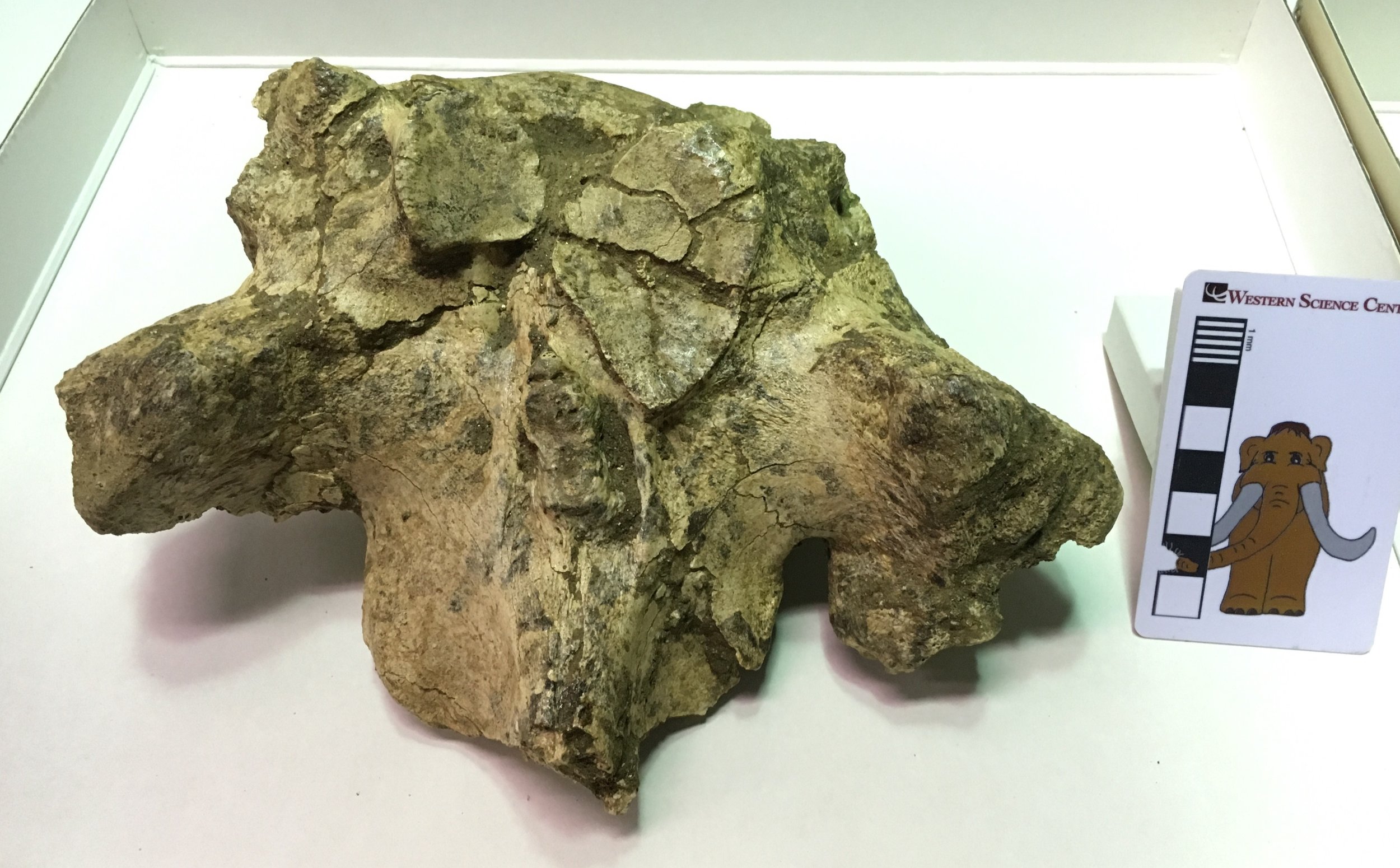 Typically, a vertebra articulates at three points with each vertebra in front of and behind it. Two of those points are the anterior and posterior surfaces of the centrum (so the anterior surface of the centrum articulates with the posterior surface of the centrum in front). The other articulations are located on the neural arch or spine. These articulations are often found at the tips of projections called zygapophyses, so the two prezygapophyses on the front edge of the neural arch will articulate with the corresponding pair of postzygapophyses on the next vertebra in front. The actual anterior articulation areas are called the prezygapophyseal articular facets (because zygapophysis was not an intimidating enough term by itself)!In mastodon thoracic vertebrae the prezygapophysis doesn't really stick out as a projection, but the articular facets themselves are very large. Below is the dorsal view image with the articular facets outlined in red:
 The left and right prezygapophyseal articular facets should be nearly mirror images of each other, but that's clearly not the case here. The right facet is more than twice as large as the left, and is actually so large that it extends slightly across the midline of the vertebra onto the left side. In addition, note the area outlined in blue, shown below in an oblique closeup:
 This is an osteophyte, a pathological bony growth that may form due to injury or as a result of osteoarthritis. It's not clear exactly what happened to this mastodon. The vertebra could have grown abnormally due to a genetic condition, and the resulting unusual stress caused by a deformed vertebra could have caused osteoarthritis later in life. Alternatively, the mastodon could have suffered an infection or injury that resulted in the deformed vertebra. The injury needn't have occurred in this spot; a muscle or bone injury elsewhere in the back or even in the legs or feet could have placed enough asymmetrical stress on the back to cause this deformation. Basically, walking with a limp over a long period of time could have potentially caused a condition like this. Since we have only a few bones from this animal, it's likely we'll never know for sure why this mastodon had such an asymmetrical vertebra.
Fossil Friday - sloth ulna
 Last Saturday the Western Science Center was visited by Eric Scott from The Cooper Center and high school student Santiago Hernandez. They came with a specific goal in mind, to look at fossil horses from a small, understudied collection, called the Harveston Collection, from western Riverside County.While the Harveston Collection is dominated by horses, there are several other kinds of animal present, including bison, camels, deer, and mammoths. As we examined each drawer looking for horses, Eric noticed the bone shown above, which was labeled in the field as a juvenile bison humerus. We both immediately agreed that this bone was not a humerus, as is lacked the distinctive articulations found in the humerus at both the proximal and distal ends. After a few minutes of debate, we decided that the bone looked more like a damaged ulna (a bone from the forearm), perhaps from a ground sloth. Conveniently, the ground sloth Paramylodon was common at Diamond Valley Lake, and we have a cast mounted skeleton on exhibit. So, with the horses temporarily forgotten, we rushed off to the exhibit hall to compare the Harveston specimen to Paramylodon (thanks to Santiago for the photo):
 It turned out that our suspicion was correct; allowing for breakage, the bone is a perfect match for the right ulna of Paramylodon. This was a neat discovery, as this is the only sloth bone currently identified from the Harveston Collection (although I think sloths had previously been found at nearby localities). There are new discoveries to be found in museum collections all over the world.
Fossil Friday - eggshell fragment
 With Easter coming up in a few days, it seems an appropriate time to feature one of the rarer fossils from Diamond Valley Lake, an eggshell fragment.While bird eggs are largely mineralized (unlike leathery lizard, snake, and turtle eggs), they are of course thin and fragile, as anyone who has dropped a carton of eggs knows. Moreover, if an egg lasts long enough to produce a baby bird, the chick shatters it upon hatching and then tramples the fragments in the nest. All this adds up to fragments of eggs, rather than complete, intact eggs. The fragment shown here is one of the largest recovered from Diamond Valley Lake. The curvature suggests that it may have come from a relatively large egg (chicken-sized, maybe), but there's a good deal of uncertainty involved in such a prediction. Most eggs are not spherical, but are instead, well, egg-shaped, which means that the curvature is different on different parts of the egg.Fossils such as eggshells also serve as a reminder of the effects taphonomic bias has on our knowledge of the fossil record. So far, 17 species of birds have been identified from Diamond Valley Lake deposits, although the actual number of bird species was undoubtedly much higher. Those deposits span tens of thousands of years (maybe as much as 200,000). How many eggs would 17 species of birds lay in 100,000 years? Yet, in the deposits we have more mastodon teeth than eggshell fragments. The eggs were mostly broken into tiny, even dust-sized fragments, and the calcium carbonate-rich shells are susceptible to dissolving in acidic groundwater. Between predators, hatching and trampling, and abiotic processes such as dissolution, what must have originally been many millions of eggs was reduced to just a handful of identifiable fragments. Taphonomic processes will selectively remove certain remains, leaving us a fossil record biased in favor of some organisms and against others. This is something to always keep in mind when examining fossil deposits.
